5YVZ

Crystal structure of the Kdo hydroxylase KdoO, a non-heme Fe(II) alphaketoglutarate dependent dioxygenase in complex with alphaketoglutarate and Fe(III)


Experimental Data Snapshot

  • Method: X-RAY DIFFRACTION
  • Resolution: 1.60 Å
  • R-Value Free: 0.182 
  • R-Value Work: 0.160 
  • R-Value Observed: 0.161 

wwPDB Validation   3D Report Full Report


This is version 1.2 of the entry. See complete history


Literature

Biochemical and Structural Insights into an Fe(II)/ alpha-Ketoglutarate/O2-Dependent Dioxygenase, Kdo 3-Hydroxylase (KdoO).

Joo, S.H.Pemble, C.W.Yang, E.G.Raetz, C.R.H.Chung, H.S.

(2018) J Mol Biol 430: 4036-4048

  • DOI: https://doi.org/10.1016/j.jmb.2018.07.029
  • Primary Citation of Related Structures:  
    5YKA, 5YVZ, 5YW0, 6A2E

  • PubMed Abstract: 

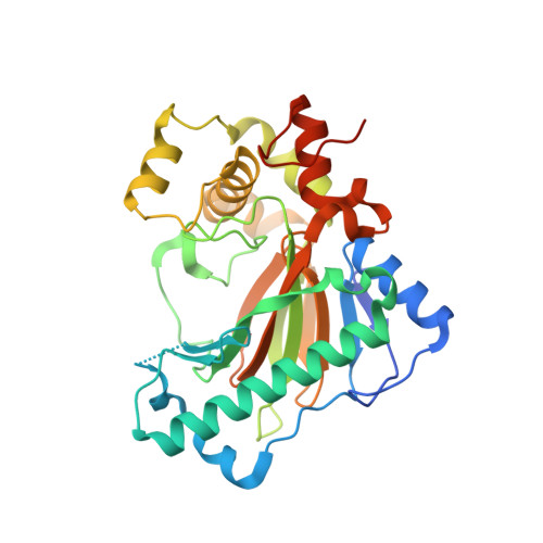
    During lipopolysaccharide biosynthesis in several pathogens, including Burkholderia and Yersinia, 3-deoxy-d-manno-oct-2-ulosonic acid (Kdo) 3-hydroxylase, otherwise referred to as KdoO, converts Kdo to d-glycero-d-talo-oct-2-ulosonic acid (Ko) in an Fe(II)/α-ketoglutarate (α-KG)/O 2 -dependent manner. This conversion renders the bacterial outer membrane more stable and resistant to stresses such as an acidic environment. KdoO is a membrane-associated, deoxy-sugar hydroxylase that does not show significant sequence identity with any known enzymes, and its structural information has not been previously reported. Here, we report the biochemical and structural characterization of KdoO, Minf_1012 (Kdo MI ), from Methylacidiphilum infernorum V4. The de novo structure of Kdo MI apoprotein indicates that KdoO MI consists of 13 α helices and 11 β strands, and has the jelly roll fold containing a metal binding motif, HXDX 111 H. Structures of Kdo MI bound to Co(II), Kdo MI bound to α-KG and Fe(III), and Kdo MI bound to succinate and Fe(III), in addition to mutagenesis analysis, indicate that His146, His260, and Asp148 play critical roles in Fe(II) binding, while Arg127, Arg162, Arg174, and Trp176 stabilize α-KG. It was also observed that His225 is adjacent to the active site and plays an important role in the catalysis of KdoO MI without affecting substrate binding, possibly being involved in oxygen activation. The crystal structure of KdoO MI is the first completed structure of a deoxy-sugar hydroxylase, and the data presented here have provided mechanistic insights into deoxy-sugar hydroxylase, KdoO, and lipopolysaccharide biosynthesis.


  • Organizational Affiliation

    Department of Biochemistry, Duke University Medical Center, Durham, NC 27710, USA; Department of Pharmacy, Daegu Catholic University, Gyeongbuk 38430, South Korea.


Macromolecules
Find similar proteins by:  (by identity cutoff)  |  3D Structure
Entity ID: 1
MoleculeChains Sequence LengthOrganismDetailsImage
Uncharacterized protein KdoO318Methylacidiphilum infernorum V4Mutation(s): 0 
Gene Names: Minf_1012
UniProt
Find proteins for B3DUR4 (Methylacidiphilum infernorum (isolate V4))
Explore B3DUR4 
Go to UniProtKB:  B3DUR4
Entity Groups  
Sequence Clusters30% Identity50% Identity70% Identity90% Identity95% Identity100% Identity
UniProt GroupB3DUR4
Sequence Annotations
Expand
  • Reference Sequence
Small Molecules
Ligands 6 Unique
IDChains Name / Formula / InChI Key2D Diagram3D Interactions
PG4
Query on PG4

Download Ideal Coordinates CCD File 
E [auth A],
F [auth A],
G [auth A],
H [auth A]
TETRAETHYLENE GLYCOL
C8 H18 O5
UWHCKJMYHZGTIT-UHFFFAOYSA-N
AKG
Query on AKG

Download Ideal Coordinates CCD File 
C [auth A]2-OXOGLUTARIC ACID
C5 H6 O5
KPGXRSRHYNQIFN-UHFFFAOYSA-N
SO4
Query on SO4

Download Ideal Coordinates CCD File 
K [auth A]SULFATE ION
O4 S
QAOWNCQODCNURD-UHFFFAOYSA-L
GOL
Query on GOL

Download Ideal Coordinates CCD File 
D [auth A]GLYCEROL
C3 H8 O3
PEDCQBHIVMGVHV-UHFFFAOYSA-N
ACT
Query on ACT

Download Ideal Coordinates CCD File 
I [auth A],
J [auth A]
ACETATE ION
C2 H3 O2
QTBSBXVTEAMEQO-UHFFFAOYSA-M
FE
Query on FE

Download Ideal Coordinates CCD File 
B [auth A]FE (III) ION
Fe
VTLYFUHAOXGGBS-UHFFFAOYSA-N
Experimental Data & Validation

Experimental Data

  • Method: X-RAY DIFFRACTION
  • Resolution: 1.60 Å
  • R-Value Free: 0.182 
  • R-Value Work: 0.160 
  • R-Value Observed: 0.161 
  • Space Group: P 21 21 21
Unit Cell:
Length ( Å )Angle ( ˚ )
a = 45.71α = 90
b = 59.204β = 90
c = 116.264γ = 90
Software Package:
Software NamePurpose
HKL-2000data scaling
PHENIXrefinement
PDB_EXTRACTdata extraction
HKL-2000data reduction
PHENIXphasing

Structure Validation

View Full Validation Report


Entry History & Funding Information

Deposition Data


Funding OrganizationLocationGrant Number
NIHUnited States--

Revision History  (Full details and data files)

  • Version 1.0: 2018-09-05
    Type: Initial release
  • Version 1.1: 2018-10-24
    Changes: Data collection, Database references
  • Version 1.2: 2023-11-22
    Changes: Advisory, Data collection, Database references, Derived calculations, Refinement description