5YVK

Crystal structure of a cyclase Famc1 from Fischerella ambigua UTEX 1903


Experimental Data Snapshot

  • Method: X-RAY DIFFRACTION
  • Resolution: 1.29 Å
  • R-Value Free: 0.187 
  • R-Value Work: 0.166 
  • R-Value Observed: 0.166 

wwPDB Validation   3D Report Full Report


Ligand Structure Quality Assessment 


This is version 1.2 of the entry. See complete history


Literature

The Crystal Structure of a Class of Cyclases that Catalyze the Cope Rearrangement

Chen, C.C.Hu, X.Tang, X.Yang, Y.Ko, T.P.Gao, J.Zheng, Y.Huang, J.W.Yu, Z.Li, L.Han, S.Cai, N.Zhang, Y.Liu, W.Guo, R.T.

(2018) Angew Chem Int Ed Engl 57: 15060-15064

  • DOI: https://doi.org/10.1002/anie.201808231
  • Primary Citation of Related Structures:  
    5YVK, 5YVL, 5YVP, 5Z53, 5Z54, 5ZFJ, 6A8X, 6A92, 6A98, 6A99, 6A9F, 6ADU

  • PubMed Abstract: 

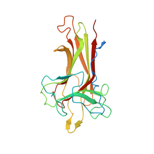
    Found recently in stignomatales, the Stig cyclases catalyze the Cope rearrangement and intramolecular cyclization to produce complex indole alkaloids. Five crystal structures were solved of subfamily 1 and 2 Stig cyclases, which adopt a β-sandwich fold like the non-catalytic carbohydrate-binding motif. Several complex structures were also determined of indole-based compounds, which are bound to the hydrophobic terminal cavity, where a conserved Asp residue makes an H-bond to the indole N and triggers the acid-catalyzed Cope rearrangement. Through analyzing the enzyme-ligand interactions and mutagenesis experiments, several aromatic residues were found important in catalysis. Apart from a common substrate binding mode and catalytic mechanism, potential subfamily variations that may attribute to the different product specificity are implicated. These results shall expand our scope of enzymology, in particular for further investigation of the biosynthetic Cope rearrangement.


  • Organizational Affiliation

    State Key Laboratory of Biocatalysis and Enzyme Engineering, Hubei Collaborative Innovation Center for Green Transformation of Bio-resources, Environmental Microbial Technology Center of Hubei Province, Hubei Key Laboratory of Industrial Biotechnology, College of Life Sciences, Hubei University, Wuhan, 430062, China.


Macromolecules
Find similar proteins by:  (by identity cutoff)  |  3D Structure
Entity ID: 1
MoleculeChains Sequence LengthOrganismDetailsImage
cyclase225Fischerella ambigua UTEX 1903Mutation(s): 0 
UniProt
Find proteins for V5TER4 (Fischerella ambigua (strain UTEX 1903))
Explore V5TER4 
Go to UniProtKB:  V5TER4
Entity Groups  
Sequence Clusters30% Identity50% Identity70% Identity90% Identity95% Identity100% Identity
UniProt GroupV5TER4
Sequence Annotations
Expand
  • Reference Sequence
Small Molecules
Ligands 2 Unique
IDChains Name / Formula / InChI Key2D Diagram3D Interactions
0HZ
Query on 0HZ

Download Ideal Coordinates CCD File 
B [auth A]amino({3-[(3S,8aS)-1,4-dioxooctahydropyrrolo[1,2-a]pyrazin-3-yl]propyl}amino)methaniminium
C11 H20 N5 O2
ZRJHYOXNWCMGMW-JGVFFNPUSA-O
CA
Query on CA

Download Ideal Coordinates CCD File 
C [auth A],
D [auth A]
CALCIUM ION
Ca
BHPQYMZQTOCNFJ-UHFFFAOYSA-N
Experimental Data & Validation

Experimental Data

  • Method: X-RAY DIFFRACTION
  • Resolution: 1.29 Å
  • R-Value Free: 0.187 
  • R-Value Work: 0.166 
  • R-Value Observed: 0.166 
  • Space Group: P 31 2 1
Unit Cell:
Length ( Å )Angle ( ˚ )
a = 54.845α = 90
b = 54.845β = 90
c = 152.562γ = 120
Software Package:
Software NamePurpose
PHENIXrefinement
HKL-2000data scaling
PDB_EXTRACTdata extraction
HKL-2000data reduction
PHENIXphasing

Structure Validation

View Full Validation Report


Ligand Structure Quality Assessment 


Entry History 

Deposition Data

Revision History  (Full details and data files)

  • Version 1.0: 2018-11-07
    Type: Initial release
  • Version 1.1: 2018-11-21
    Changes: Data collection, Database references
  • Version 1.2: 2018-12-26
    Changes: Data collection, Structure summary