


Crystal structure of a novel KAS III from Acinetobacter baumannii
Lee, W.C., Jung, M., Lee, J., Kim, Y.To be published.
Experimental Data Snapshot
Entity ID: 1 | |||||
|---|---|---|---|---|---|
| Molecule | Chains | Sequence Length | Organism | Details | Image |
| 3-Oxoacyl-[acyl-carrier-(ACP)] synthase III C terminal family protein | 371 | Acinetobacter baumannii | Mutation(s): 1 Gene Names: KAS III EC: 2.3.1.180 (PDB Primary Data), 2.3.1.41 (PDB Primary Data) | 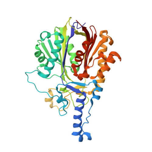 | |
UniProt | |||||
Find proteins for V5VGF8 (Acinetobacter baumannii) Explore V5VGF8 Go to UniProtKB: V5VGF8 | |||||
Entity Groups | |||||
| Sequence Clusters | 30% Identity50% Identity70% Identity90% Identity95% Identity100% Identity | ||||
| UniProt Group | V5VGF8 | ||||
Sequence AnnotationsExpand | |||||
| |||||
| Ligands 2 Unique | |||||
|---|---|---|---|---|---|
| ID | Chains | Name / Formula / InChI Key | 2D Diagram | 3D Interactions | |
| COA Query on COA | F [auth B] | COENZYME A C21 H36 N7 O16 P3 S RGJOEKWQDUBAIZ-IBOSZNHHSA-N |  | ||
| GOL Query on GOL | C [auth A], D [auth B], E [auth B] | GLYCEROL C3 H8 O3 PEDCQBHIVMGVHV-UHFFFAOYSA-N |  | ||
| Length ( Å ) | Angle ( ˚ ) |
|---|---|
| a = 75.135 | α = 90 |
| b = 106.942 | β = 90 |
| c = 109.959 | γ = 90 |
| Software Name | Purpose |
|---|---|
| SCALEPACK | data scaling |
| PHENIX | refinement |
| PDB_EXTRACT | data extraction |
| HKL-2000 | data reduction |
| PHASER | phasing |
| Funding Organization | Location | Grant Number |
|---|---|---|
| National Research Foundation of Korea | Korea, Republic Of | MSIP-2016R1A2B2008543 |
RCSB PDB (citation) is hosted by
RCSB PDB is a member of the

