5YHF

Crystal structure of SecDF in Super-membrane-facing form


Experimental Data Snapshot

  • Method: X-RAY DIFFRACTION
  • Resolution: 2.80 Å
  • R-Value Free: 0.246 
  • R-Value Work: 0.203 
  • R-Value Observed: 0.207 

wwPDB Validation   3D Report Full Report


Ligand Structure Quality Assessment 


This is version 1.3 of the entry. See complete history


Literature

Remote Coupled Drastic beta-Barrel to beta-Sheet Transition of the Protein Translocation Motor.

Furukawa, A.Nakayama, S.Yoshikaie, K.Tanaka, Y.Tsukazaki, T.

(2018) Structure 26: 485-489.e2

  • DOI: https://doi.org/10.1016/j.str.2018.01.002
  • Primary Citation of Related Structures:  
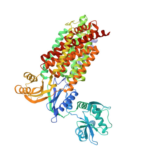
    5YHF

  • PubMed Abstract: 

    The membrane protein SecDF, belonging to the RND superfamily, enhances protein translocation at the extracytoplasmic side using a proton gradient. Here, we report the crystal structure of SecDF in a form we named Super-membrane-facing (Super F) form, demonstrating a β-barrel architecture instead of the previously reported β-sheet structure. Through this structural insight and supporting results of an in vivo crosslinking experiment, we propose a remote coupling model in which a structural change of the transmembrane region drives a functional, extracytoplasmic conformational transition.


  • Organizational Affiliation

    Graduate School of Biological Sciences, Nara Institute of Science and Technology, 8916-5 Takayama-cho, Ikoma, Nara 630-0192, Japan.


Macromolecules
Find similar proteins by:  (by identity cutoff)  |  3D Structure
Entity ID: 1
MoleculeChains Sequence LengthOrganismDetailsImage
Protein translocase subunit SecDF741Thermus thermophilus HB8Mutation(s): 1 
Gene Names: secDFTTHA0697
Membrane Entity: Yes 
UniProt
Find proteins for Q5SKE6 (Thermus thermophilus (strain ATCC 27634 / DSM 579 / HB8))
Explore Q5SKE6 
Go to UniProtKB:  Q5SKE6
Entity Groups  
Sequence Clusters30% Identity50% Identity70% Identity90% Identity95% Identity100% Identity
UniProt GroupQ5SKE6
Sequence Annotations
Expand
  • Reference Sequence
Experimental Data & Validation

Experimental Data

  • Method: X-RAY DIFFRACTION
  • Resolution: 2.80 Å
  • R-Value Free: 0.246 
  • R-Value Work: 0.203 
  • R-Value Observed: 0.207 
  • Space Group: P 1
Unit Cell:
Length ( Å )Angle ( ˚ )
a = 55.19α = 75.11
b = 65.64β = 75.23
c = 79.98γ = 80.83
Software Package:
Software NamePurpose
PHENIXrefinement
XSCALEdata scaling
MoRDaphasing
PDB_EXTRACTdata extraction
XDSdata collection

Structure Validation

View Full Validation Report


Ligand Structure Quality Assessment 


Entry History & Funding Information

Deposition Data


Funding OrganizationLocationGrant Number
JSPS/MEXT KAKENHIJapanJP26119007, JP26291023, JP17H05669, JP16K14713, JP15H01537, JP15J08235, 15K14490, JP15K06972
Mitsubishi FoundationJapan--
Noguchi InstituteJapan--
Naito FoundationJapan--
Mochida Memorial Foundation for Medical and Pharmaceutical ResearchJapan--

Revision History  (Full details and data files)

  • Version 1.0: 2018-02-14
    Type: Initial release
  • Version 1.1: 2018-02-21
    Changes: Database references, Structure summary
  • Version 1.2: 2018-03-21
    Changes: Database references
  • Version 1.3: 2023-11-22
    Changes: Data collection, Database references, Refinement description