5Y8T

Crystal structure of Bacillus subtilis PadR in complex with p-coumaric acid


Experimental Data Snapshot

  • Method: X-RAY DIFFRACTION
  • Resolution: 2.00 Å
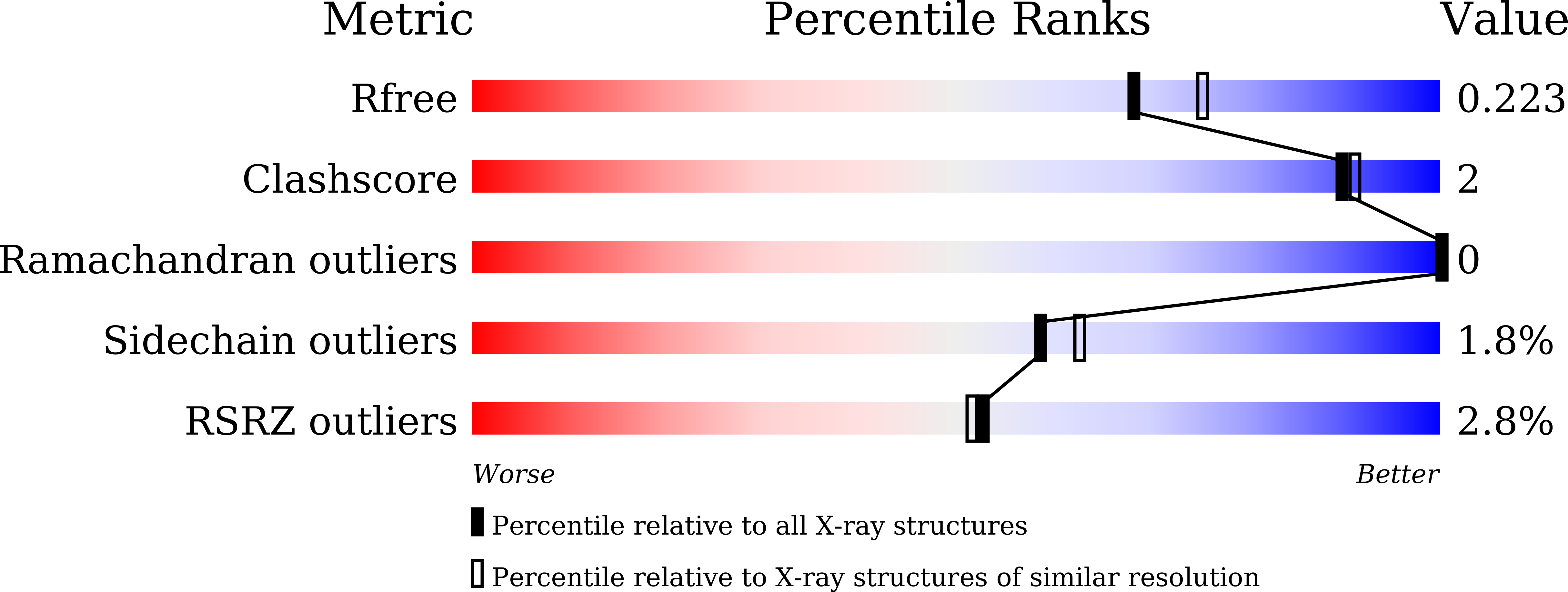
  • R-Value Free: 0.222 
  • R-Value Work: 0.195 
  • R-Value Observed: 0.197 

wwPDB Validation   3D Report Full Report


Ligand Structure Quality Assessment 


This is version 1.2 of the entry. See complete history


Literature

Structural basis of effector and operator recognition by the phenolic acid-responsive transcriptional regulator PadR.

Park, S.C.Kwak, Y.M.Song, W.S.Hong, M.Yoon, S.I.

(2017) Nucleic Acids Res 45: 13080-13093

  • DOI: https://doi.org/10.1093/nar/gkx1055
  • Primary Citation of Related Structures:  
    5X11, 5X12, 5X13, 5X14, 5Y8T

  • PubMed Abstract: 

    The PadR family is a large group of transcriptional regulators that function as environmental sensors. PadR negatively controls the expression of phenolic acid decarboxylase, which detoxifies harmful phenolic acids. To identify the mechanism by which PadR regulates phenolic acid-mediated gene expression, we performed structural and mutational studies of effector and operator recognition by Bacillus subtilis PadR. PadR contains an N-terminal winged helix-turn-helix (wHTH) domain (NTD) and a C-terminal homodimerization domain (CTD) and dimerizes into a dolmen shape. The PadR dimer interacts with the palindromic sequence of the operator DNA using the NTD. Two tyrosine residues and a positively charged residue in the NTD provide major DNA-binding energy and are highly conserved in the PadR family, suggesting that these three residues represent the canonical DNA-binding motif of the PadR family. PadR directly binds a phenolic acid effector molecule using a unique interdomain pocket created between the NTD and the CTD. Although the effector-binding site of PadR is positionally segregated from the DNA-binding site, effector binding to the interdomain pocket causes PadR to be rearranged into a DNA binding-incompatible conformer through an allosteric interdomain-reorganization mechanism.


  • Organizational Affiliation

    Division of Biomedical Convergence, College of Biomedical Science, Kangwon National University, Chuncheon 24341, Republic of Korea.


Macromolecules
Find similar proteins by:  (by identity cutoff)  |  3D Structure
Entity ID: 1
MoleculeChains Sequence LengthOrganismDetailsImage
Transcriptional regulator
A, B, C
188Bacillus spizizenii str. W23Mutation(s): 0 
Gene Names: padRBSUW23_04210
UniProt
Find proteins for E0TW95 (Bacillus spizizenii (strain ATCC 23059 / NRRL B-14472 / W23))
Explore E0TW95 
Go to UniProtKB:  E0TW95
Entity Groups  
Sequence Clusters30% Identity50% Identity70% Identity90% Identity95% Identity100% Identity
UniProt GroupE0TW95
Sequence Annotations
Expand
  • Reference Sequence
Small Molecules
Binding Affinity Annotations 
IDSourceBinding Affinity
HC4 Binding MOAD:  5Y8T Kd: 6200 (nM) from 1 assay(s)
Experimental Data & Validation

Experimental Data

  • Method: X-RAY DIFFRACTION
  • Resolution: 2.00 Å
  • R-Value Free: 0.222 
  • R-Value Work: 0.195 
  • R-Value Observed: 0.197 
  • Space Group: C 1 2 1
Unit Cell:
Length ( Å )Angle ( ˚ )
a = 93.217α = 90
b = 125.301β = 113.8
c = 79.657γ = 90
Software Package:
Software NamePurpose
REFMACrefinement
HKL-2000data reduction
HKL-2000data scaling
PHASERphasing

Structure Validation

View Full Validation Report


Ligand Structure Quality Assessment 


Entry History 

Deposition Data

Revision History  (Full details and data files)

  • Version 1.0: 2017-11-29
    Type: Initial release
  • Version 1.1: 2017-12-27
    Changes: Database references
  • Version 1.2: 2023-11-22
    Changes: Data collection, Database references, Refinement description