5Y3Z

Structure of the periplasmic domain of the MotB L119P mutant from Salmonella (crystal form 1)


Experimental Data Snapshot

  • Method: X-RAY DIFFRACTION
  • Resolution: 2.00 Å
  • R-Value Free: 0.221 
  • R-Value Work: 0.190 
  • R-Value Observed: 0.193 

wwPDB Validation   3D Report Full Report


Ligand Structure Quality Assessment 


This is version 1.2 of the entry. See complete history


Literature

The Helix Rearrangement in the Periplasmic Domain of the Flagellar Stator B Subunit Activates Peptidoglycan Binding and Ion Influx.

Kojima, S.Takao, M.Almira, G.Kawahara, I.Sakuma, M.Homma, M.Kojima, C.Imada, K.

(2018) Structure 26: 590-598.e5

  • DOI: https://doi.org/10.1016/j.str.2018.02.016
  • Primary Citation of Related Structures:  
    5Y3Z, 5Y40

  • PubMed Abstract: 

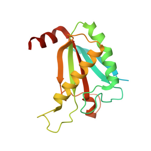
    The stator of the bacterial flagellar motor couples ion flow with torque generation. The ion-conducting stator channel opens only when incorporated into and anchored around the rotor via the peptidoglycan (PG) binding domain of the B subunit (MotB C ). However, no direct evidence of PG binding coupled with channel activation has been presented. Here, we report the structural rearrangements of MotB C responsible for this coupling process. A MotB C fragment with the L119P replacement, which is known to cause channel activation, was able to bind PG. Nuclear magnetic resonance analysis of MotB C and the crystal structure of the MotB C -L119P dimer revealed major structural changes in helix α1. In vivo crosslinking results confirm that a major rearrangement occurs. Our results suggest that, upon stator incorporation into the motor, helix α1 of MotB C changes into an extended non-helical structure. We propose that this change allows the stator both to bind PG and to open its proton channel.


  • Organizational Affiliation

    Division of Biological Science, Graduate School of Science, Nagoya University, Chikusa-Ku, Nagoya 464-8602, Japan. Electronic address: z47616a@cc.nagoya-u.ac.jp.


Macromolecules
Find similar proteins by:  (by identity cutoff)  |  3D Structure
Entity ID: 1
MoleculeChains Sequence LengthOrganismDetailsImage
Motility protein B
A, B
183Salmonella enterica subsp. enterica serovar Typhimurium str. LT2Mutation(s): 1 
Gene Names: motBSTM1922
UniProt
Find proteins for P55892 (Salmonella typhimurium (strain LT2 / SGSC1412 / ATCC 700720))
Explore P55892 
Go to UniProtKB:  P55892
Entity Groups  
Sequence Clusters30% Identity50% Identity70% Identity90% Identity95% Identity100% Identity
UniProt GroupP55892
Sequence Annotations
Expand
  • Reference Sequence
Small Molecules
Ligands 3 Unique
IDChains Name / Formula / InChI Key2D Diagram3D Interactions
ARG
Query on ARG

Download Ideal Coordinates CCD File 
C [auth A],
D [auth A],
H [auth B]
ARGININE
C6 H15 N4 O2
ODKSFYDXXFIFQN-BYPYZUCNSA-O
MPD
Query on MPD

Download Ideal Coordinates CCD File 
F [auth A],
G [auth A],
J [auth B]
(4S)-2-METHYL-2,4-PENTANEDIOL
C6 H14 O2
SVTBMSDMJJWYQN-YFKPBYRVSA-N
IMD
Query on IMD

Download Ideal Coordinates CCD File 
E [auth A],
I [auth B]
IMIDAZOLE
C3 H5 N2
RAXXELZNTBOGNW-UHFFFAOYSA-O
Experimental Data & Validation

Experimental Data

  • Method: X-RAY DIFFRACTION
  • Resolution: 2.00 Å
  • R-Value Free: 0.221 
  • R-Value Work: 0.190 
  • R-Value Observed: 0.193 
  • Space Group: P 21 21 21
Unit Cell:
Length ( Å )Angle ( ˚ )
a = 44.13α = 90
b = 84.8β = 90
c = 86.83γ = 90
Software Package:
Software NamePurpose
PHENIXrefinement
iMOSFLMdata reduction
Aimlessdata scaling
PHENIXphasing

Structure Validation

View Full Validation Report


Ligand Structure Quality Assessment 


Entry History & Funding Information

Deposition Data


Funding OrganizationLocationGrant Number
Japan--

Revision History  (Full details and data files)

  • Version 1.0: 2018-04-11
    Type: Initial release
  • Version 1.1: 2018-04-18
    Changes: Data collection, Database references
  • Version 1.2: 2023-11-22
    Changes: Data collection, Database references, Refinement description