


Crystal structure of ycgT from bacillus subtilis
Komori, H.To be published.
Experimental Data Snapshot
Entity ID: 1 | |||||
|---|---|---|---|---|---|
| Molecule | Chains | Sequence Length | Organism | Details | Image |
| Ferredoxin--NADP reductase | 329 | Bacillus subtilis | Mutation(s): 0 Gene Names: B4417_2052 EC: 1.18.1.2 | 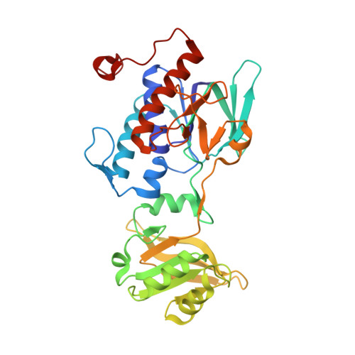 | |
UniProt | |||||
Find proteins for O31475 (Bacillus subtilis (strain 168)) Explore O31475 Go to UniProtKB: O31475 | |||||
Entity Groups | |||||
| Sequence Clusters | 30% Identity50% Identity70% Identity90% Identity95% Identity100% Identity | ||||
| UniProt Group | O31475 | ||||
Sequence AnnotationsExpand | |||||
| |||||
| Ligands 1 Unique | |||||
|---|---|---|---|---|---|
| ID | Chains | Name / Formula / InChI Key | 2D Diagram | 3D Interactions | |
| FAD Query on FAD | B [auth A] | FLAVIN-ADENINE DINUCLEOTIDE C27 H33 N9 O15 P2 VWWQXMAJTJZDQX-UYBVJOGSSA-N |  | ||
| Length ( Å ) | Angle ( ˚ ) |
|---|---|
| a = 72.371 | α = 90 |
| b = 164.598 | β = 90 |
| c = 76.576 | γ = 90 |
| Software Name | Purpose |
|---|---|
| HKL-2000 | data scaling |
| REFMAC | refinement |
| PDB_EXTRACT | data extraction |
| PHASER | phasing |
| HKL | data scaling |
RCSB PDB (citation) is hosted by
RCSB PDB is a member of the

