5XFV

Crystal structures of FMN-bound form of dihydroorotate dehydrogenase from Trypanosoma brucei


Experimental Data Snapshot

  • Method: X-RAY DIFFRACTION
  • Resolution: 1.79 Å
  • R-Value Free: 0.198 
  • R-Value Work: 0.166 
  • R-Value Observed: 0.168 

wwPDB Validation   3D Report Full Report


Ligand Structure Quality Assessment 


This is version 1.1 of the entry. See complete history


Literature

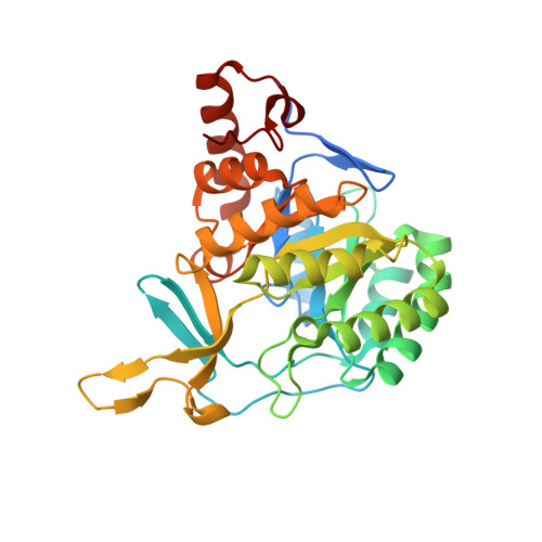
Crystal structures of FMN-bound and FMN-free forms of dihydroorotate dehydrogenase fromTrypanosoma brucei.

Kubota, T.Tani, O.Yamaguchi, T.Namatame, I.Sakashita, H.Furukawa, K.Yamasaki, K.

(2018) FEBS Open Bio 8: 680-691

  • DOI: https://doi.org/10.1002/2211-5463.12403
  • Primary Citation of Related Structures:  
    5XFV, 5XFW

  • PubMed Abstract: 

    Dihydroorotate dehydrogenase (DHODH) is a flavin-binding enzyme essential for pyrimidine biosynthesis, which converts dihydroorotate to orotate. Three-dimensional structures of cytosolic DHODH of parasitic protozoa are of interest in drug discovery for neglected tropical diseases, especially because these enzymes possess significantly different structural and functional properties from the membrane-associated human enzyme. The existing crystal structures of the flavin mononucleotide (FMN)-bound DHODHs reveal a number of interactions stabilizing FMN. However, to understand the binding mechanism correctly, it is necessary to compare the structures of the FMN-bound and FMN-free forms, because the protein moiety of the former is not necessarily the same as the latter. Here, we prepared the FMN-free DHODH of Trypanosoma brucei using an Escherichia coli overexpression system. Although this apoform lacks enzymatic activity, simple incubation with FMN activated the enzyme. It was stable enough to be crystallized, enabling us to determine its structure by X-ray crystallography at 1.6 Å resolution. We also determined the FMN-bound form at 1.8 Å resolution. Although the two structures have essentially the same scaffold, we observed flipping of a peptide-bond plane in the vicinity of the FMN-binding site, accompanied by an alternative hydrogen-bonding pattern. Comparisons of B factors of the protein main chain revealed that binding of FMN decreased flexibility of most of the residues at the FMN-binding site, but increased flexibility of a lid-like loop structure over the active center. This increase was ascribed to a conformational change in an FMN-contacting residue, Asn195, which induced a rearrangement of a hydrogen-bond network of the residues comprising the lid.


  • Organizational Affiliation

    Biomedical Research Institute National Institute of Advanced Industrial Science and Technology (AIST) Tsukuba Japan.


Macromolecules
Find similar proteins by:  (by identity cutoff)  |  3D Structure
Entity ID: 1
MoleculeChains Sequence LengthOrganismDetailsImage
Dihydroorotate dehydrogenase (fumarate)
A, B, C, D
334Trypanosoma brucei brucei TREU927Mutation(s): 1 
Gene Names: Tb927.5.3830
EC: 1.3.98.1
UniProt
Find proteins for Q57U83 (Trypanosoma brucei brucei (strain 927/4 GUTat10.1))
Explore Q57U83 
Go to UniProtKB:  Q57U83
Entity Groups  
Sequence Clusters30% Identity50% Identity70% Identity90% Identity95% Identity100% Identity
UniProt GroupQ57U83
Sequence Annotations
Expand
  • Reference Sequence
Small Molecules
Ligands 2 Unique
IDChains Name / Formula / InChI Key2D Diagram3D Interactions
FMN
Query on FMN

Download Ideal Coordinates CCD File 
E [auth A],
I [auth B],
M [auth C],
R [auth D]
FLAVIN MONONUCLEOTIDE
C17 H21 N4 O9 P
FVTCRASFADXXNN-SCRDCRAPSA-N
MLI
Query on MLI

Download Ideal Coordinates CCD File 
F [auth A]
G [auth A]
H [auth A]
J [auth B]
K [auth B]
F [auth A],
G [auth A],
H [auth A],
J [auth B],
K [auth B],
L [auth B],
N [auth C],
O [auth C],
P [auth C],
Q [auth C],
S [auth D]
MALONATE ION
C3 H2 O4
OFOBLEOULBTSOW-UHFFFAOYSA-L
Binding Affinity Annotations 
IDSourceBinding Affinity
FMN Binding MOAD:  5XFV Kd: 1.20e-5 (nM) from 1 assay(s)
Experimental Data & Validation

Experimental Data

  • Method: X-RAY DIFFRACTION
  • Resolution: 1.79 Å
  • R-Value Free: 0.198 
  • R-Value Work: 0.166 
  • R-Value Observed: 0.168 
  • Space Group: P 21 21 2
Unit Cell:
Length ( Å )Angle ( ˚ )
a = 139.914α = 90
b = 146.979β = 90
c = 66.491γ = 90
Software Package:
Software NamePurpose
REFMACrefinement
HKL-2000data reduction
HKL-2000data scaling
PHASERphasing

Structure Validation

View Full Validation Report


Ligand Structure Quality Assessment 


Entry History 

Deposition Data

Revision History  (Full details and data files)

  • Version 1.0: 2018-04-25
    Type: Initial release
  • Version 1.1: 2023-11-22
    Changes: Data collection, Database references, Refinement description