5XBP

Oxygenase component of 3-nitrotoluene dioxygenase from Diaphorobacter sp. strain DS2


Experimental Data Snapshot

  • Method: X-RAY DIFFRACTION
  • Resolution: 2.90 Å
  • R-Value Free: 0.284 
  • R-Value Work: 0.241 
  • R-Value Observed: 0.241 

wwPDB Validation   3D Report Full Report


This is version 1.2 of the entry. See complete history


Literature

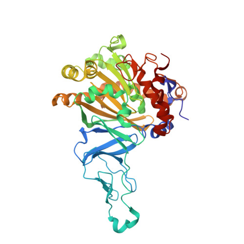
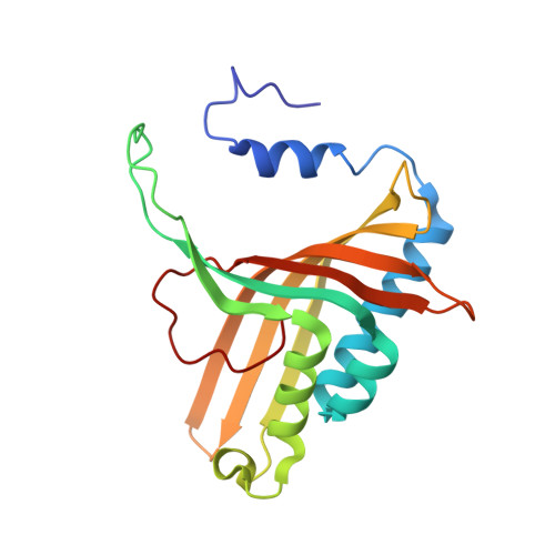
Structural and functional studies of ferredoxin and oxygenase components of 3-nitrotoluene dioxygenase from Diaphorobacter sp. strain DS2.

Kumari, A.Singh, D.Ramaswamy, S.Ramanathan, G.

(2017) PLoS One 12: e0176398-e0176398

  • DOI: https://doi.org/10.1371/journal.pone.0176398
  • Primary Citation of Related Structures:  
    5XBP

  • PubMed Abstract: 

    3-nitrotoluene dioxygenase (3NTDO) from Diaphorobacter sp. strain DS2 catalyses the conversion of 3-nitrotoluene (3NT) into a mixture of 3- and 4-methylcatechols with release of nitrite. We report here, X-ray crystal structures of oxygenase and ferredoxin components of 3NTDO at 2.9 Å and 2.4 Å, respectively. The residues responsible for nitrite release in 3NTDO were further probed by four single and two double mutations in the catalytic site of α-subunit of the dioxygenase. Modification of Val 350 to Phe, Ile 204 to Ala, and Asn258 to Val by site directed mutagenesis resulted in inactive enzymes revealing the importance of these residues in catalysis. Docking studies of meta nitrotoluene to the active site of 3NTDO suggested possible orientations of binding that favor the formation of 3-methylcatechol (3MC) over 4-methylcatechol energetically. The electron transfer pathway from ferredoxin subunit to the active site of the oxygenase subunit is also proposed.


  • Organizational Affiliation

    Department of Chemistry, Indian Institute of Technology Kanpur, Kalyanpur, Kanpur, Uttar Pradesh, India.


Macromolecules
Find similar proteins by:  (by identity cutoff)  |  3D Structure
Entity ID: 1
MoleculeChains Sequence LengthOrganismDetailsImage
3NT oxygenase alpha subunitA,
C [auth D],
E [auth G]
446Diaphorobacter sp. DS2Mutation(s): 0 
Gene Names: mntAc
UniProt
Find proteins for M9PW10 (Diaphorobacter sp. DS2)
Explore M9PW10 
Go to UniProtKB:  M9PW10
Entity Groups  
Sequence Clusters30% Identity50% Identity70% Identity90% Identity95% Identity100% Identity
UniProt GroupM9PW10
Sequence Annotations
Expand
  • Reference Sequence
Find similar proteins by:  (by identity cutoff)  |  3D Structure
Entity ID: 2
MoleculeChains Sequence LengthOrganismDetailsImage
3NT oxygenase beta subunitB [auth C],
D [auth F],
F [auth I]
193Diaphorobacter sp. DS2Mutation(s): 0 
Gene Names: mntAd
UniProt
Find proteins for M9PV03 (Diaphorobacter sp. DS2)
Explore M9PV03 
Go to UniProtKB:  M9PV03
Entity Groups  
Sequence Clusters30% Identity50% Identity70% Identity90% Identity95% Identity100% Identity
UniProt GroupM9PV03
Sequence Annotations
Expand
  • Reference Sequence
Experimental Data & Validation

Experimental Data

  • Method: X-RAY DIFFRACTION
  • Resolution: 2.90 Å
  • R-Value Free: 0.284 
  • R-Value Work: 0.241 
  • R-Value Observed: 0.241 
  • Space Group: P 31 2 1
Unit Cell:
Length ( Å )Angle ( ˚ )
a = 178.804α = 90
b = 178.804β = 90
c = 242.323γ = 120
Software Package:
Software NamePurpose
PHENIXrefinement
SCALAdata scaling
XDSdata reduction
PHASERphasing

Structure Validation

View Full Validation Report


Entry History & Funding Information

Deposition Data


Funding OrganizationLocationGrant Number
Department of biotechnologyIndiaDBT/PR5801/INF/22/156/2012

Revision History  (Full details and data files)

  • Version 1.0: 2017-04-19
    Type: Initial release
  • Version 1.1: 2018-02-21
    Changes: Database references
  • Version 1.2: 2023-11-22
    Changes: Data collection, Database references, Derived calculations, Refinement description