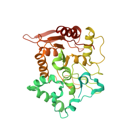
Crystal structure of mimivirus uracil-DNA glycosylase
Kwon, E., Pathak, D., Chang, H.W., Kim, D.Y.(2017) PLoS One 12: e0182382-e0182382
- PubMed: 28763516
- DOI: https://doi.org/10.1371/journal.pone.0182382
- Primary Citation of Related Structures:
5X55 - PubMed Abstract:
Cytosine deamination induced by stresses or enzymatic catalysis converts deoxycytidine into deoxyuridine, thereby introducing a G to A mutation after DNA replication. Base-excision repair to correct uracil to cytosine is initiated by uracil-DNA glycosylase (UDG), which recognizes and eliminates uracil from DNA. Mimivirus, one of the largest known viruses, also encodes a distinctive UDG gene containing a long N-terminal domain (N-domain; residues 1-130) and a motif-I (residues 327-343), in addition to the canonical catalytic domain of family I UDGs (also called UNGs). To understand the structural and functional features of the additional segments, we have determined the crystal structure of UNG from Acanthamoeba polyphaga mimivirus (mvUNG). In the crystal structure of mvUNG, residues 95-130 in the N-domain bind to a hydrophobic groove in the catalytic domain, and motif-I forms a short β-sheet with a positively charged surface near the active site. Circular dichroism spectra showed that residues 1-94 are in a random coil conformation. Deletion of the three additional fragments reduced the activity and thermal stability, compared to full-length mvUNG. The results suggested that the mvUNG N-domain and motif-I are required for its structural and functional integrity.
Organizational Affiliation:
College of Pharmacy, Yeungnam University, Gyeongsan, Gyeongbuk, South Korea.