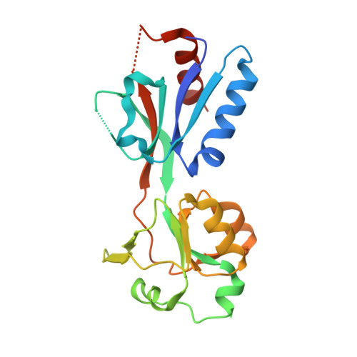
Crystal Structure of the Regulatory Domain of AphB from Vibrio vulnificus, a Virulence Gene Regulator
Park, N., Song, S., Choi, G., Jang, K.K., Jo, I., Choi, S.H., Ha, N.-C.(2017) Mol Cells 40: 299-306
- PubMed: 28427249
- DOI: https://doi.org/10.14348/molcells.2017.0015
- Primary Citation of Related Structures:
5FHK, 5X0N, 5X0O - PubMed Abstract:
The transcriptional activator AphB has been implicated in acid resistance and pathogenesis in the food borne pathogens Vibrio vulnificus and Vibrio cholerae . To date, the full-length AphB crystal structure of V. cholerae has been determined and characterized by a tetrameric assembly of AphB consisting of a DNA binding domain and a regulatory domain (RD). Although acidic pH and low oxygen tension might be involved in the activation of AphB, it remains unknown which ligand or stimulus activates AphB at the molecular level. In this study, we determine the crystal structure of the AphB RD from V. vulnificus under aerobic conditions without modification at the conserved cysteine residue of the RD, even in the presence of the oxidizing agent cumene hydroperoxide. A cysteine to serine amino acid residue mutant RD protein further confirmed that the cysteine residue is not involved in sensing oxidative stress in vitro. Interestingly, an unidentified small molecule was observed in the inter-subdomain cavity in the RD when the crystal was incubated with cumene hydroperoxide molecules, suggesting a new ligand-binding site. In addition, we confirmed the role of AphB in acid tolerance by observing an aphB -dependent increase in cadC transcript level when V. vulnificus was exposed to acidic pH. Our study contributes to the understanding of the AphB molecular mechanism in the process of recognizing the host environment.
Organizational Affiliation:
Department of Agricultural Biotechnology, Center for Food Safety and Toxicology, and Research Institute for Agriculture and Life Sciences, Seoul National University, Seoul 08826, Korea.