5WUK

Crystal structure of EED [G255D] in complex with EZH2 peptide and EED226 compound


Experimental Data Snapshot

  • Method: X-RAY DIFFRACTION
  • Resolution: 2.03 Å
  • R-Value Free: 0.191 
  • R-Value Work: 0.160 
  • R-Value Observed: 0.162 

wwPDB Validation   3D Report Full Report


Ligand Structure Quality Assessment 


This is version 1.0 of the entry. See complete history


Literature

Split luciferase-based biosensors for characterizing EED binders

Li, L.Feng, L.Shi, M.Zeng, J.Chen, Z.Zhong, L.Huang, L.Guo, W.Huang, Y.Qi, W.Lu, C.Li, E.Zhao, K.Gu, J.

(2017) Anal Biochem 522: 37-45

  • DOI: https://doi.org/10.1016/j.ab.2017.01.014
  • Primary Citation of Related Structures:  
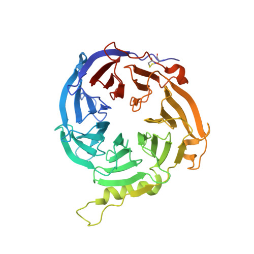
    5WUK

  • PubMed Abstract: 

    The EED (embryonic ectoderm development) subunit of the Polycomb repressive complex 2 (PRC2) plays an important role in the feed forward regulation of the PRC2 enzymatic activity. We recently identified a new class of allosteric PRC2 inhibitors that bind to the H3K27me3 pocket of EED. Multiple assays were developed and used to identify and characterize this type of PRC2 inhibitors. One of them is a genetically encoded EED biosensor based on the EED[G255D] mutant and the split firefly luciferase. This EED biosensor can detect the compound binding in the transfected cells and in the in vitro biochemical assays. Compared to other commonly used cellular assays, the EED biosensor assay has the advantage of shorter compound incubation with cells. The in vitro EED biosensor is much more sensitive than other label-free biophysical assays (e.g. DSF, ITC). Based on the crystal structure, the DSF data as well as the biosensor assay data, it's most likely that compound-induced increase in the luciferase activity of the EED[G255D] biosensor results from the decreased non-productive interactions between the EED subdomain and other subdomains within the biosensor construct. This new insight of the mechanism might help to broaden the use of the split luciferase based biosensors.


  • Organizational Affiliation

    China Novartis Institutes for Biomedical Research, 4218 Jinke Road, Shanghai 201203, China. Electronic address: ling-1.li@novartis.com.


Macromolecules
Find similar proteins by:  (by identity cutoff)  |  3D Structure
Entity ID: 1
MoleculeChains Sequence LengthOrganismDetailsImage
Polycomb protein EED366Homo sapiensMutation(s): 1 
Gene Names: EED
UniProt & NIH Common Fund Data Resources
Find proteins for O75530 (Homo sapiens)
Explore O75530 
Go to UniProtKB:  O75530
PHAROS:  O75530
GTEx:  ENSG00000074266 
Entity Groups  
Sequence Clusters30% Identity50% Identity70% Identity90% Identity95% Identity100% Identity
UniProt GroupO75530
Sequence Annotations
Expand
  • Reference Sequence
Find similar proteins by:  (by identity cutoff)  |  3D Structure
Entity ID: 2
MoleculeChains Sequence LengthOrganismDetailsImage
Histone-lysine N-methyltransferase EZH228Homo sapiensMutation(s): 0 
EC: 2.1.1.43
UniProt & NIH Common Fund Data Resources
Find proteins for Q15910 (Homo sapiens)
Explore Q15910 
Go to UniProtKB:  Q15910
PHAROS:  Q15910
GTEx:  ENSG00000106462 
Entity Groups  
Sequence Clusters30% Identity50% Identity70% Identity90% Identity95% Identity100% Identity
UniProt GroupQ15910
Sequence Annotations
Expand
  • Reference Sequence
Small Molecules
Ligands 2 Unique
IDChains Name / Formula / InChI Key2D Diagram3D Interactions
73K
Query on 73K

Download Ideal Coordinates CCD File 
D [auth A]N-(furan-2-ylmethyl)-8-(4-methylsulfonylphenyl)-[1,2,4]triazolo[4,3-c]pyrimidin-5-amine
C17 H15 N5 O3 S
DYIRSNMPIZZNBK-UHFFFAOYSA-N
GOL
Query on GOL

Download Ideal Coordinates CCD File 
C [auth A]GLYCEROL
C3 H8 O3
PEDCQBHIVMGVHV-UHFFFAOYSA-N
Binding Affinity Annotations 
IDSourceBinding Affinity
73K BindingDB:  5WUK Kd: min: 82, max: 114 (nM) from 2 assay(s)
IC50: min: 18, max: 220 (nM) from 9 assay(s)
Binding MOAD:  5WUK Kd: 14 (nM) from 1 assay(s)
Experimental Data & Validation

Experimental Data

  • Method: X-RAY DIFFRACTION
  • Resolution: 2.03 Å
  • R-Value Free: 0.191 
  • R-Value Work: 0.160 
  • R-Value Observed: 0.162 
  • Space Group: P 21 21 21
Unit Cell:
Length ( Å )Angle ( ˚ )
a = 53.1α = 90
b = 80.28β = 90
c = 108.42γ = 90
Software Package:
Software NamePurpose
SCALEPACKdata scaling
BUSTERrefinement
PDB_EXTRACTdata extraction

Structure Validation

View Full Validation Report


Ligand Structure Quality Assessment 


Entry History 

Deposition Data

  • Released Date: 2017-05-03 
  • Deposition Author(s): Chen, Z.

Revision History  (Full details and data files)

  • Version 1.0: 2017-05-03
    Type: Initial release