5WIK

JAK2 Pseudokinase in complex with BI-D1870


Experimental Data Snapshot

  • Method: X-RAY DIFFRACTION
  • Resolution: 2.60 Å
  • R-Value Free: 0.251 
  • R-Value Work: 0.206 
  • R-Value Observed: 0.211 

wwPDB Validation   3D Report Full Report


Ligand Structure Quality Assessment 


This is version 1.3 of the entry. See complete history


Literature

Discovery and Structural Characterization of ATP-Site Ligands for the Wild-Type and V617F Mutant JAK2 Pseudokinase Domain.

McNally, R.Li, Q.Li, K.Dekker, C.Vangrevelinghe, E.Jones, M.Chene, P.Machauer, R.Radimerski, T.Eck, M.J.

(2019) ACS Chem Biol 14: 587-593

  • DOI: https://doi.org/10.1021/acschembio.8b00722
  • Primary Citation of Related Structures:  
    5WIJ, 5WIK, 5WIL, 5WIM, 5WIN, 6D2I, 6G3C

  • PubMed Abstract: 

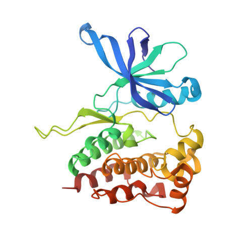
    The oncogenic V617F mutation lies in the pseudokinase domain of JAK2, marking it as a potential target for development of compounds that might inhibit the pathogenic activity of the mutant protein. We used differential scanning fluorimetry to identify compounds that bind the JAK2 pseudokinase domain. Crystal structures of five candidate compounds with the wild-type domain reveal their modes of binding. Exploration of analogs of screening hit BI-D1870 led to the identification of compound 2, a 123 nM ligand for the pseudokinase domain. Interestingly, crystal structures of the V617F domain in complex with two unrelated compounds reveal a conformation that is characteristic of the wild-type domain, rather than that previously observed for the V617F mutant. These structures suggest that certain ATP-site ligands can modulate the V617F allosteric site, thereby providing a mechanistic rationale for targeting the pseudokinase domain and a structural foundation for development of more potent and pseudokinase-selective compounds.


  • Organizational Affiliation

    Department of Biological Chemistry and Molecular Pharmacology, Harvard Medical School and Department of Cancer Biology , Dana-Farber Cancer Institute , Boston , Massachusetts 02115 , United States.


Macromolecules
Find similar proteins by:  (by identity cutoff)  |  3D Structure
Entity ID: 1
MoleculeChains Sequence LengthOrganismDetailsImage
Tyrosine-protein kinase JAK2A [auth B]277Homo sapiensMutation(s): 3 
Gene Names: JAK2
EC: 2.7.10.2
UniProt & NIH Common Fund Data Resources
Find proteins for O60674 (Homo sapiens)
Explore O60674 
Go to UniProtKB:  O60674
PHAROS:  O60674
GTEx:  ENSG00000096968 
Entity Groups  
Sequence Clusters30% Identity50% Identity70% Identity90% Identity95% Identity100% Identity
UniProt GroupO60674
Sequence Annotations
Expand
  • Reference Sequence
Small Molecules
Ligands 1 Unique
IDChains Name / Formula / InChI Key2D Diagram3D Interactions
584
Query on 584

Download Ideal Coordinates CCD File 
B
(7R)-2-[(3,5-difluoro-4-hydroxyphenyl)amino]-5,7-dimethyl-8-(3-methylbutyl)-7,8-dihydropteridin-6(5H)-one
C19 H23 F2 N5 O2
DTEKTGDVSARYDS-LLVKDONJSA-N
Experimental Data & Validation

Experimental Data

  • Method: X-RAY DIFFRACTION
  • Resolution: 2.60 Å
  • R-Value Free: 0.251 
  • R-Value Work: 0.206 
  • R-Value Observed: 0.211 
  • Space Group: P 21 21 21
Unit Cell:
Length ( Å )Angle ( ˚ )
a = 57.125α = 90
b = 60.844β = 90
c = 89.724γ = 90
Software Package:
Software NamePurpose
PHENIXrefinement
xia2data reduction
Aimlessdata scaling
PHASERphasing

Structure Validation

View Full Validation Report


Ligand Structure Quality Assessment 


Entry History 

Deposition Data

Revision History  (Full details and data files)

  • Version 1.0: 2018-08-01
    Type: Initial release
  • Version 1.1: 2019-03-20
    Changes: Data collection, Database references
  • Version 1.2: 2019-05-01
    Changes: Data collection, Database references
  • Version 1.3: 2024-03-13
    Changes: Data collection, Database references