5WGL

Crystal structure of Danio rerio histone deacetylase 6 catalytic domain 2 in complex with ricolinostat (ACY-1215)


Experimental Data Snapshot

  • Method: X-RAY DIFFRACTION
  • Resolution: 1.70 Å
  • R-Value Free: 0.209 
  • R-Value Work: 0.184 
  • R-Value Observed: 0.185 

wwPDB Validation   3D Report Full Report


Ligand Structure Quality Assessment 


This is version 1.3 of the entry. See complete history


Literature

Unusual zinc-binding mode of HDAC6-selective hydroxamate inhibitors.

Porter, N.J.Mahendran, A.Breslow, R.Christianson, D.W.

(2017) Proc Natl Acad Sci U S A 114: 13459-13464

  • DOI: https://doi.org/10.1073/pnas.1718823114
  • Primary Citation of Related Structures:  
    5WGI, 5WGK, 5WGL, 5WGM

  • PubMed Abstract: 

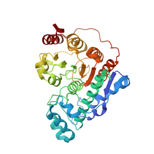
    Histone deacetylases (HDACs) regulate myriad cellular processes by catalyzing the hydrolysis of acetyl-l-lysine residues in histone and nonhistone proteins. The Zn 2+ -dependent class IIb enzyme HDAC6 regulates microtubule function by deacetylating α-tubulin, which suppresses microtubule dynamics and leads to cell cycle arrest and apoptosis. Accordingly, HDAC6 is a target for the development of selective inhibitors that might be useful in new therapeutic approaches for the treatment of cancer, neurodegenerative diseases, and other disorders. Here, we present high-resolution structures of catalytic domain 2 from Danio rerio HDAC6 (henceforth simply "HDAC6") complexed with compounds that selectively inhibit HDAC6 while maintaining nanomolar inhibitory potency: N -hydroxy-4-[( N (2-hydroxyethyl)-2-phenylacetamido)methyl)-benzamide)] (HPB), ACY-1215 (Ricolinostat), and ACY-1083. These structures reveal that an unusual monodentate Zn 2+ coordination mode is exploited by sterically bulky HDAC6-selective phenylhydroxamate inhibitors. We additionally report the ultrahigh-resolution structure of the HDAC6-trichostatin A complex, which reveals two Zn 2+ -binding conformers for the inhibitor: a major conformer (70%) with canonical bidentate hydroxamate-Zn 2+ coordination geometry and a minor conformer (30%) with monodentate hydroxamate-Zn 2+ coordination geometry, reflecting a free energy difference of only 0.5 kcal/mol. The minor conformer is not visible in lower resolution structure determinations. Structural comparisons of HDAC6-inhibitor complexes with class I HDACs suggest active site features that contribute to the isozyme selectivity observed in biochemical assays.


  • Organizational Affiliation

    Roy and Diana Vagelos Laboratories, Department of Chemistry, University of Pennsylvania, Philadelphia, PA 19104-6323.


Macromolecules
Find similar proteins by:  (by identity cutoff)  |  3D Structure
Entity ID: 1
MoleculeChains Sequence LengthOrganismDetailsImage
Hdac6 protein
A, B
364Danio rerioMutation(s): 0 
Gene Names: hdac6
UniProt
Find proteins for F8W4B7 (Danio rerio)
Explore F8W4B7 
Go to UniProtKB:  F8W4B7
Entity Groups  
Sequence Clusters30% Identity50% Identity70% Identity90% Identity95% Identity100% Identity
UniProt GroupF8W4B7
Sequence Annotations
Expand
  • Reference Sequence
Small Molecules
Ligands 6 Unique
IDChains Name / Formula / InChI Key2D Diagram3D Interactions
AH4
Query on AH4

Download Ideal Coordinates CCD File 
F [auth A],
S [auth B]
2-(diphenylamino)-N-[7-(hydroxyamino)-7-oxoheptyl]pyrimidine-5-carboxamide
C24 H27 N5 O3
QGZYDVAGYRLSKP-UHFFFAOYSA-N
FLC
Query on FLC

Download Ideal Coordinates CCD File 
O [auth A]CITRATE ANION
C6 H5 O7
KRKNYBCHXYNGOX-UHFFFAOYSA-K
PEG
Query on PEG

Download Ideal Coordinates CCD File 
N [auth A]DI(HYDROXYETHYL)ETHER
C4 H10 O3
MTHSVFCYNBDYFN-UHFFFAOYSA-N
ZN
Query on ZN

Download Ideal Coordinates CCD File 
C [auth A],
P [auth B]
ZINC ION
Zn
PTFCDOFLOPIGGS-UHFFFAOYSA-N
EDO
Query on EDO

Download Ideal Coordinates CCD File 
AA [auth B]
BA [auth B]
G [auth A]
H [auth A]
I [auth A]
AA [auth B],
BA [auth B],
G [auth A],
H [auth A],
I [auth A],
J [auth A],
K [auth A],
L [auth A],
M [auth A],
T [auth B],
U [auth B],
V [auth B],
W [auth B],
X [auth B],
Y [auth B],
Z [auth B]
1,2-ETHANEDIOL
C2 H6 O2
LYCAIKOWRPUZTN-UHFFFAOYSA-N
K
Query on K

Download Ideal Coordinates CCD File 
D [auth A],
E [auth A],
Q [auth B],
R [auth B]
POTASSIUM ION
K
NPYPAHLBTDXSSS-UHFFFAOYSA-N
Experimental Data & Validation

Experimental Data

  • Method: X-RAY DIFFRACTION
  • Resolution: 1.70 Å
  • R-Value Free: 0.209 
  • R-Value Work: 0.184 
  • R-Value Observed: 0.185 
  • Space Group: P 21 21 21
Unit Cell:
Length ( Å )Angle ( ˚ )
a = 87.189α = 90
b = 88.002β = 90
c = 119.025γ = 90
Software Package:
Software NamePurpose
PHENIXrefinement
iMOSFLMdata reduction
Aimlessdata scaling
PHASERphasing

Structure Validation

View Full Validation Report


Ligand Structure Quality Assessment 


Entry History 

Deposition Data

Revision History  (Full details and data files)

  • Version 1.0: 2017-12-06
    Type: Initial release
  • Version 1.1: 2017-12-20
    Changes: Database references
  • Version 1.2: 2018-01-03
    Changes: Database references
  • Version 1.3: 2023-10-04
    Changes: Data collection, Database references, Derived calculations, Refinement description