5VZU

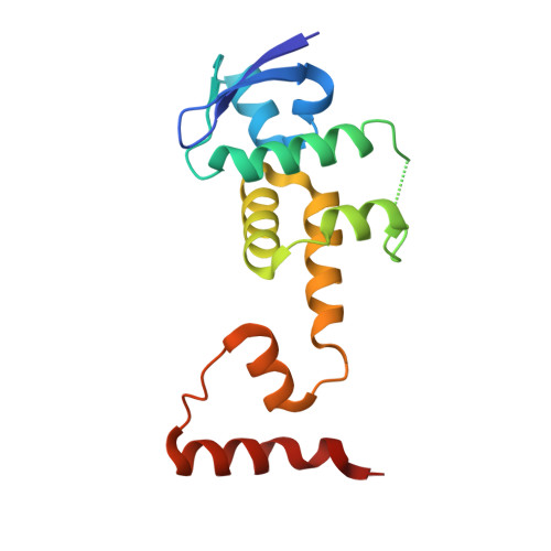
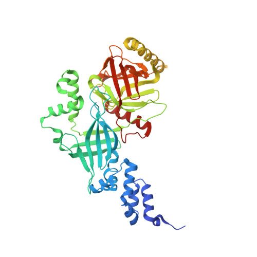
Crystal structure of the Skp1-FBXO31-cyclin D1 complex

  • Classification: CELL CYCLE
  • Organism(s): Homo sapiens
  • Expression System: Escherichia coli BL21(DE3)
  • Mutation(s): Yes 

  • Deposited: 2017-05-29 Released: 2018-01-17 
  • Deposition Author(s): Li, Y., Jin, K., Hao, B.
  • Funding Organization(s): National Institutes of Health/National Institute of General Medical Sciences (NIH/NIGMS), Connecticut Regenerative Medicine Research Fund

Experimental Data Snapshot

  • Method: X-RAY DIFFRACTION
  • Resolution: 2.70 Å
  • R-Value Free: 0.233 
  • R-Value Work: 0.180 
  • R-Value Observed: 0.183 

wwPDB Validation   3D Report Full Report


This is version 1.2 of the entry. See complete history


Literature

Structural basis of the phosphorylation-independent recognition of cyclin D1 by the SCFFBXO31 ubiquitin ligase.

Li, Y.Jin, K.Bunker, E.Zhang, X.Luo, X.Liu, X.Hao, B.

(2018) Proc Natl Acad Sci U S A 115: 319-324

  • DOI: https://doi.org/10.1073/pnas.1708677115
  • Primary Citation of Related Structures:  
    5VZT, 5VZU

  • PubMed Abstract: 

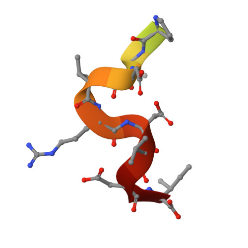
    Ubiquitin-dependent proteolysis of cyclin D1 is associated with normal and tumor cell proliferation and survival. The SCF FBXO31 (Skp1-Cul1-Rbx1-FBXO31) ubiquitin ligase complex mediates genotoxic stress-induced cyclin D1 degradation. Previous studies have suggested that cyclin D1 levels are maintained at steady state by phosphorylation-dependent nuclear export and subsequent proteolysis in the cytoplasm. Here we present the crystal structures of the Skp1-FBXO31 complex alone and bound to a phosphorylated cyclin D1 C-terminal peptide. FBXO31 possesses a unique substrate-binding domain consisting of two β-barrel motifs, whereas cyclin D1 binds to FBXO31 by tucking its free C-terminal carboxylate tail into an open cavity of the C-terminal FBXO31 β-barrel. Biophysical and functional studies demonstrate that SCF FBXO31 is capable of recruiting and ubiquitinating cyclin D1 in a phosphorylation-independent manner. Our findings provide a conceptual framework for understanding the substrate specificity of the F-box protein FBXO31 and the mechanism of FBXO31-regulated cyclin D1 protein turnover.


  • Organizational Affiliation

    Department of Molecular Biology and Biophysics, University of Connecticut Health Center, Farmington, CT 06030.


Macromolecules
Find similar proteins by:  (by identity cutoff)  |  3D Structure
Entity ID: 1
MoleculeChains Sequence LengthOrganismDetailsImage
S-phase kinase-associated protein 1
A, C
149Homo sapiensMutation(s): 1 
Gene Names: SKP1EMC19OCP2SKP1ATCEB1L
UniProt & NIH Common Fund Data Resources
Find proteins for P63208 (Homo sapiens)
Explore P63208 
Go to UniProtKB:  P63208
PHAROS:  P63208
GTEx:  ENSG00000113558 
Entity Groups  
Sequence Clusters30% Identity50% Identity70% Identity90% Identity95% Identity100% Identity
UniProt GroupP63208
Sequence Annotations
Expand
  • Reference Sequence
Find similar proteins by:  (by identity cutoff)  |  3D Structure
Entity ID: 2
MoleculeChains Sequence LengthOrganismDetailsImage
F-box only protein 31
B, D
488Homo sapiensMutation(s): 0 
Gene Names: FBXO31FBX14FBX31PP2386
UniProt & NIH Common Fund Data Resources
Find proteins for Q5XUX0 (Homo sapiens)
Explore Q5XUX0 
Go to UniProtKB:  Q5XUX0
PHAROS:  Q5XUX0
GTEx:  ENSG00000103264 
Entity Groups  
Sequence Clusters30% Identity50% Identity70% Identity90% Identity95% Identity100% Identity
UniProt GroupQ5XUX0
Sequence Annotations
Expand
  • Reference Sequence

Find similar proteins by:  Sequence   |   3D Structure  

Entity ID: 3
MoleculeChains Sequence LengthOrganismDetailsImage
Cyclin D1
E, F
17Homo sapiensMutation(s): 0 
UniProt & NIH Common Fund Data Resources
Find proteins for P24385 (Homo sapiens)
Explore P24385 
Go to UniProtKB:  P24385
PHAROS:  P24385
GTEx:  ENSG00000110092 
Entity Groups  
Sequence Clusters30% Identity50% Identity70% Identity90% Identity95% Identity100% Identity
UniProt GroupP24385
Sequence Annotations
Expand
  • Reference Sequence
Small Molecules
Ligands 2 Unique
IDChains Name / Formula / InChI Key2D Diagram3D Interactions
PO4
Query on PO4

Download Ideal Coordinates CCD File 
H [auth B]
I [auth B]
J [auth B]
K [auth B]
L [auth B]
H [auth B],
I [auth B],
J [auth B],
K [auth B],
L [auth B],
N [auth D],
O [auth D],
P [auth D],
Q [auth D],
R [auth D]
PHOSPHATE ION
O4 P
NBIIXXVUZAFLBC-UHFFFAOYSA-K
ZN
Query on ZN

Download Ideal Coordinates CCD File 
G [auth B],
M [auth D]
ZINC ION
Zn
PTFCDOFLOPIGGS-UHFFFAOYSA-N
Modified Residues  1 Unique
IDChains TypeFormula2D DiagramParent
TPO
Query on TPO
E, F
L-PEPTIDE LINKINGC4 H10 N O6 PTHR
Experimental Data & Validation

Experimental Data

  • Method: X-RAY DIFFRACTION
  • Resolution: 2.70 Å
  • R-Value Free: 0.233 
  • R-Value Work: 0.180 
  • R-Value Observed: 0.183 
  • Space Group: C 1 2 1
Unit Cell:
Length ( Å )Angle ( ˚ )
a = 87.679α = 90
b = 156.235β = 104.21
c = 155.522γ = 90
Software Package:
Software NamePurpose
REFMACrefinement
HKL-2000data scaling
PDB_EXTRACTdata extraction
HKL-2000data reduction
PHASERphasing

Structure Validation

View Full Validation Report


Entry History & Funding Information

Deposition Data


Funding OrganizationLocationGrant Number
National Institutes of Health/National Institute of General Medical Sciences (NIH/NIGMS)United StatesGM099948
Connecticut Regenerative Medicine Research FundUnited States15-RMA-UCHC-04

Revision History  (Full details and data files)

  • Version 1.0: 2018-01-17
    Type: Initial release
  • Version 1.1: 2020-01-01
    Changes: Author supporting evidence
  • Version 1.2: 2023-10-04
    Changes: Data collection, Database references, Refinement description