5VCL

Structure of the Qdm peptide bound to Qa-1a


Experimental Data Snapshot

  • Method: X-RAY DIFFRACTION
  • Resolution: 2.05 Å
  • R-Value Free: 0.246 
  • R-Value Work: 0.217 
  • R-Value Observed: 0.218 

wwPDB Validation   3D Report Full Report


This is version 1.1 of the entry. See complete history


Literature

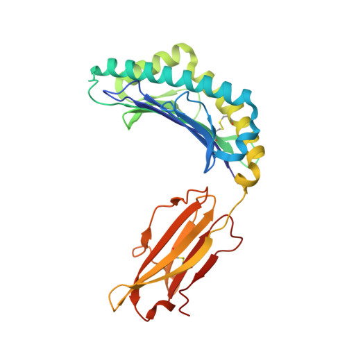
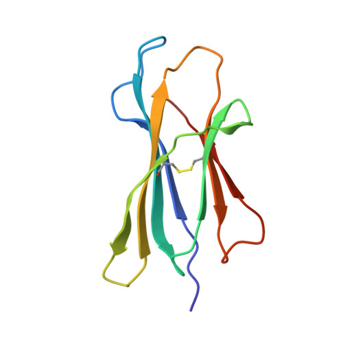
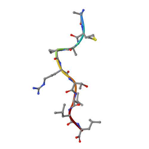
Crystal structure of Qa-1a with bound Qa-1 determinant modifier peptide.

Ying, G.Wang, J.Kumar, V.Zajonc, D.M.

(2017) PLoS One 12: e0182296-e0182296

  • DOI: https://doi.org/10.1371/journal.pone.0182296
  • Primary Citation of Related Structures:  
    5VCL

  • PubMed Abstract: 

    Qa-1 is a non-classical Major Histocompatibility (MHC) class I molecule that generally presents hydrophobic peptides including Qdm derived from the leader sequence of classical MHC I molecules for immune surveillance by NK cells. Qa-1 bound peptides derived from the TCR Vβ8.2 of activated T cells also activates CD8+ regulatory T cells to control autoimmunity and maintain self-tolerance. Four allotypes of Qa-1 (Qa-1a-d) are expressed that are highly conserved in sequence but have several variations that could affect peptide binding to Qa-1 or TCR recognition. Here, we determined the structure of Qa-1a with bound Qdm peptide. While the overall structure is very similar to that of Qa-1b, there are several amino acid differences around the peptide binding platform that could affect TCR recognition. Most notably, two amino acid substitutions are found in the pocket P2, which binds the anchor residue Met2 of the Qdm peptide. These residues affect both the size and shape of the binding pocket, as well as affect the charge at physiologic pH, suggesting Qa-1a and Qa-1b could present slightly distinct peptide reservoirs, which could presumably be recognized by different populations of CD8+ T cells.


  • Organizational Affiliation

    Division of Cell Biology, La Jolla Institute for Allergy and Immunology (LJI), La Jolla, California, United States of America.


Macromolecules
Find similar proteins by:  (by identity cutoff)  |  3D Structure
Entity ID: 1
MoleculeChains Sequence LengthOrganismDetailsImage
H2-T23 protein278Mus musculusMutation(s): 0 
Gene Names: H2-T23
UniProt
Find proteins for Q31153 (Mus musculus)
Explore Q31153 
Go to UniProtKB:  Q31153
Entity Groups  
Sequence Clusters30% Identity50% Identity70% Identity90% Identity95% Identity100% Identity
UniProt GroupQ31153
Sequence Annotations
Expand
  • Reference Sequence
Find similar proteins by:  (by identity cutoff)  |  3D Structure
Entity ID: 2
MoleculeChains Sequence LengthOrganismDetailsImage
Beta-2-microglobulin99Mus musculusMutation(s): 0 
Gene Names: B2m
UniProt & NIH Common Fund Data Resources
Find proteins for P01887 (Mus musculus)
Explore P01887 
Go to UniProtKB:  P01887
IMPC:  MGI:88127
Entity Groups  
Sequence Clusters30% Identity50% Identity70% Identity90% Identity95% Identity100% Identity
UniProt GroupP01887
Sequence Annotations
Expand
  • Reference Sequence

Find similar proteins by:  Sequence   |   3D Structure  

Entity ID: 3
MoleculeChains Sequence LengthOrganismDetailsImage
Qdm peptideC [auth P]9Mus musculusMutation(s): 0 
UniProt
Find proteins for P01897 (Mus musculus)
Explore P01897 
Go to UniProtKB:  P01897
Entity Groups  
UniProt GroupP01897
Sequence Annotations
Expand
  • Reference Sequence
Small Molecules
Ligands 2 Unique
IDChains Name / Formula / InChI Key2D Diagram3D Interactions
GOL
Query on GOL

Download Ideal Coordinates CCD File 
D [auth A],
E [auth A],
F [auth A],
M [auth B]
GLYCEROL
C3 H8 O3
PEDCQBHIVMGVHV-UHFFFAOYSA-N
NA
Query on NA

Download Ideal Coordinates CCD File 
G [auth A]
H [auth A]
I [auth A]
J [auth A]
K [auth A]
G [auth A],
H [auth A],
I [auth A],
J [auth A],
K [auth A],
L [auth A]
SODIUM ION
Na
FKNQFGJONOIPTF-UHFFFAOYSA-N
Experimental Data & Validation

Experimental Data

  • Method: X-RAY DIFFRACTION
  • Resolution: 2.05 Å
  • R-Value Free: 0.246 
  • R-Value Work: 0.217 
  • R-Value Observed: 0.218 
  • Space Group: P 21 2 21
Unit Cell:
Length ( Å )Angle ( ˚ )
a = 67.207α = 90
b = 75.876β = 90
c = 105.211γ = 90
Software Package:
Software NamePurpose
REFMACrefinement
HKL-2000data reduction
SCALEPACKdata scaling
PHASERphasing
PDB_EXTRACTdata extraction

Structure Validation

View Full Validation Report


Entry History 

Deposition Data

Revision History  (Full details and data files)

  • Version 1.0: 2017-10-25
    Type: Initial release
  • Version 1.1: 2023-10-04
    Changes: Data collection, Database references, Derived calculations, Refinement description