5UX4

Crystal Structure of Rat Cathepsin D with (5S)-3-(5,6-dihydro-2H-pyran-3-yl)-1-fluoro- 7-(2-fluoropyridin-3-yl)spiro[chromeno[2,3- c]pyridine-5,4'-[1,3]oxazol]-2'-amine


Experimental Data Snapshot

  • Method: X-RAY DIFFRACTION
  • Resolution: 2.81 Å
  • R-Value Free: 0.237 
  • R-Value Work: 0.191 
  • R-Value Observed: 0.194 

wwPDB Validation   3D Report Full Report


Ligand Structure Quality Assessment 


This is version 2.1 of the entry. See complete history


Literature

Development of 2-aminooxazoline 3-azaxanthene beta-amyloid cleaving enzyme (BACE) inhibitors with improved selectivity against Cathepsin D.

Low, J.D.Bartberger, M.D.Chen, K.Cheng, Y.Fielden, M.R.Gore, V.Hickman, D.Liu, Q.Allen Sickmier, E.Vargas, H.M.Werner, J.White, R.D.Whittington, D.A.Wood, S.Minatti, A.E.

(2017) Medchemcomm 8: 1196-1206

  • DOI: https://doi.org/10.1039/c7md00106a
  • Primary Citation of Related Structures:  
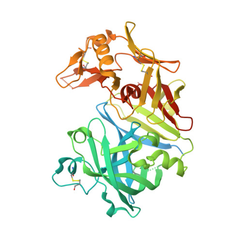
    5UX4, 5UYU

  • PubMed Abstract: 

    As part of an ongoing effort at Amgen to develop a disease-modifying therapy for Alzheimer's disease, we have previously used the aminooxazoline xanthene (AOX) scaffold to generate potent and orally efficacious BACE1 inhibitors. While AOX-BACE1 inhibitors demonstrated acceptable cardiovascular safety margins, a retinal pathological finding in rat toxicological studies demanded further investigation. It has been widely postulated that such retinal toxicity might be related to off-target inhibition of Cathepsin D (CatD), a closely related aspartyl protease. We report the development of AOX-BACE1 inhibitors with improved selectivity against CatD by following a structure- and property-based approach. Our efforts culminated in the discovery of a picolinamide-substituted 3-aza-AOX-BACE1 inhibitor absent of retinal effects in an early screening rat toxicology study.


  • Organizational Affiliation

    Department of Medicinal Chemistry , Amgen Inc. , One Amgen Center Drive , Thousand Oaks , CA 91320 , USA . Email: aminatti@amgen.com ; Tel: +1 805 447 4721.


Macromolecules
Find similar proteins by:  (by identity cutoff)  |  3D Structure
Entity ID: 1
MoleculeChains Sequence LengthOrganismDetailsImage
Cathepsin D
A, B
420Rattus norvegicusMutation(s): 0 
Gene Names: Ctsd
EC: 3.4.23.5
UniProt
Find proteins for P24268 (Rattus norvegicus)
Explore P24268 
Go to UniProtKB:  P24268
Entity Groups  
Sequence Clusters30% Identity50% Identity70% Identity90% Identity95% Identity100% Identity
UniProt GroupP24268
Sequence Annotations
Expand
  • Reference Sequence
Oligosaccharides

Help

Entity ID: 2
MoleculeChains Length2D Diagram Glycosylation3D Interactions
2-acetamido-2-deoxy-beta-D-glucopyranose-(1-4)-2-acetamido-2-deoxy-beta-D-glucopyranose
C
2N-Glycosylation
Glycosylation Resources
GlyTouCan:  G42666HT
GlyCosmos:  G42666HT
GlyGen:  G42666HT
Experimental Data & Validation

Experimental Data

  • Method: X-RAY DIFFRACTION
  • Resolution: 2.81 Å
  • R-Value Free: 0.237 
  • R-Value Work: 0.191 
  • R-Value Observed: 0.194 
  • Space Group: C 1 2 1
Unit Cell:
Length ( Å )Angle ( ˚ )
a = 136.514α = 90
b = 66.122β = 99.53
c = 99.555γ = 90
Software Package:
Software NamePurpose
HKL-2000data collection
SCALEPACKdata scaling
PHASERphasing
PHENIXrefinement
PDB_EXTRACTdata extraction
HKL-2000data reduction

Structure Validation

View Full Validation Report


Ligand Structure Quality Assessment 


Entry History 

Deposition Data

  • Released Date: 2018-06-13 
  • Deposition Author(s): Sickmier, A.

Revision History  (Full details and data files)

  • Version 1.0: 2018-06-13
    Type: Initial release
  • Version 1.1: 2018-08-29
    Changes: Data collection, Database references
  • Version 2.0: 2020-07-29
    Type: Remediation
    Reason: Carbohydrate remediation
    Changes: Atomic model, Data collection, Derived calculations, Structure summary
  • Version 2.1: 2023-10-04
    Changes: Data collection, Database references, Refinement description, Structure summary