5UQE

Multidomain structure of human kidney-type glutaminase(KGA/GLS)


Experimental Data Snapshot

  • Method: X-RAY DIFFRACTION
  • Resolution: 3.60 Å
  • R-Value Free: 0.313 
  • R-Value Work: 0.274 
  • R-Value Observed: 0.276 

wwPDB Validation   3D Report Full Report


Ligand Structure Quality Assessment 


This is version 1.5 of the entry. See complete history


Literature

The origin and evolution of human glutaminases and their atypical C-terminal ankyrin repeats.

Pasquali, C.C.Islam, Z.Adamoski, D.Ferreira, I.M.Righeto, R.D.Bettini, J.Portugal, R.V.Yue, W.W.Gonzalez, A.Dias, S.M.G.Ambrosio, A.L.B.

(2017) J Biol Chem 292: 11572-11585

  • DOI: https://doi.org/10.1074/jbc.M117.787291
  • Primary Citation of Related Structures:  
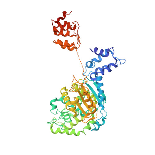
    5U0I, 5U0J, 5U0K, 5UQE

  • PubMed Abstract: 

    On the basis of tissue-specific enzyme activity and inhibition by catalytic products, Hans Krebs first demonstrated the existence of multiple glutaminases in mammals. Currently, two human genes are known to encode at least four glutaminase isoforms. However, the phylogeny of these medically relevant enzymes remains unclear, prompting us to investigate their origin and evolution. Using prokaryotic and eukaryotic glutaminase sequences, we built a phylogenetic tree whose topology suggested that the multidomain architecture was inherited from bacterial ancestors, probably simultaneously with the hosting of the proto-mitochondrion endosymbiont. We propose an evolutionary model wherein the appearance of the most active enzyme isoform, glutaminase C (GAC), which is expressed in many cancers, was a late retrotransposition event that occurred in fishes from the Chondrichthyes class. The ankyrin (ANK) repeats in the glutaminases were acquired early in their evolution. To obtain information on ANK folding, we solved two high-resolution structures of the ANK repeat-containing C termini of both kidney-type glutaminase (KGA) and GLS2 isoforms (glutaminase B and liver-type glutaminase). We found that the glutaminase ANK repeats form unique intramolecular contacts through two highly conserved motifs; curiously, this arrangement occludes a region usually involved in ANK-mediated protein-protein interactions. We also solved the crystal structure of full-length KGA and present a small-angle X-ray scattering model for full-length GLS2. These structures explain these proteins' compromised ability to assemble into catalytically active supra-tetrameric filaments, as previously shown for GAC. Collectively, these results provide information about glutaminases that may aid in the design of isoform-specific glutaminase inhibitors.


  • Organizational Affiliation

    From the Laboratório Nacional de Biociências, Centro Nacional de Pesquisa em Energia e Materiais, Campinas, São Paulo 13083-970, Brazil.


Macromolecules
Find similar proteins by:  (by identity cutoff)  |  3D Structure
Entity ID: 1
MoleculeChains Sequence LengthOrganismDetailsImage
Glutaminase kidney isoform, mitochondrialA,
B,
C,
D,
E [auth F]
520Homo sapiensMutation(s): 0 
Gene Names: GLSGLS1KIAA0838
EC: 3.5.1.2
UniProt & NIH Common Fund Data Resources
Find proteins for O94925 (Homo sapiens)
Explore O94925 
Go to UniProtKB:  O94925
PHAROS:  O94925
GTEx:  ENSG00000115419 
Entity Groups  
Sequence Clusters30% Identity50% Identity70% Identity90% Identity95% Identity100% Identity
UniProt GroupO94925
Sequence Annotations
Expand
  • Reference Sequence
Small Molecules
Ligands 1 Unique
IDChains Name / Formula / InChI Key2D Diagram3D Interactions
04A
Query on 04A

Download Ideal Coordinates CCD File 
F [auth A],
G [auth B],
H [auth F]
N,N'-[sulfanediylbis(ethane-2,1-diyl-1,3,4-thiadiazole-5,2-diyl)]bis(2-phenylacetamide)
C24 H24 N6 O2 S3
MDJIPXYRSZHCFS-UHFFFAOYSA-N
Binding Affinity Annotations 
IDSourceBinding Affinity
04A BindingDB:  5UQE Kd: min: 200, max: 4300 (nM) from 2 assay(s)
IC50: min: 25, max: 8600 (nM) from 12 assay(s)
Experimental Data & Validation

Experimental Data

  • Method: X-RAY DIFFRACTION
  • Resolution: 3.60 Å
  • R-Value Free: 0.313 
  • R-Value Work: 0.274 
  • R-Value Observed: 0.276 
  • Space Group: P 42 21 2
Unit Cell:
Length ( Å )Angle ( ˚ )
a = 144.42α = 90
b = 144.42β = 90
c = 615.22γ = 90
Software Package:
Software NamePurpose
PHENIXrefinement
MOSFLMdata reduction
Aimlessdata scaling
PHASERphasing
PDB_EXTRACTdata extraction

Structure Validation

View Full Validation Report


Ligand Structure Quality Assessment 


Entry History & Funding Information

Deposition Data


Funding OrganizationLocationGrant Number
Sao Paulo Research Foundation (FAPESP)Brazil2012/20673-2

Revision History  (Full details and data files)

  • Version 1.0: 2017-05-24
    Type: Initial release
  • Version 1.1: 2017-06-07
    Changes: Database references, Structure summary
  • Version 1.2: 2019-02-06
    Changes: Data collection, Database references
  • Version 1.3: 2019-04-17
    Changes: Author supporting evidence, Data collection
  • Version 1.4: 2020-01-01
    Changes: Author supporting evidence
  • Version 1.5: 2024-03-06
    Changes: Data collection, Database references