5UK5

Complex of Notch1(EGF8-12) bound to Jagged1(N-EGF3)

  • Classification: SIGNALING PROTEIN
  • Organism(s): Rattus norvegicus
  • Expression System: Trichoplusia ni
  • Mutation(s): Yes 

  • Deposited: 2017-01-19 Released: 2017-03-08 
  • Deposition Author(s): Garcia, K.C., Luca, V.C.
  • Funding Organization(s): National Institutes of Health/National Institute of General Medical Sciences (NIH/NIGMS), Howard Hughes Medical Institute (HHMI), Ludwig Cancer Foundation

Experimental Data Snapshot

  • Method: X-RAY DIFFRACTION
  • Resolution: 2.51 Å
  • R-Value Free: 0.263 
  • R-Value Work: 0.219 
  • R-Value Observed: 0.221 

wwPDB Validation   3D Report Full Report


Ligand Structure Quality Assessment 


This is version 2.1 of the entry. See complete history


Literature

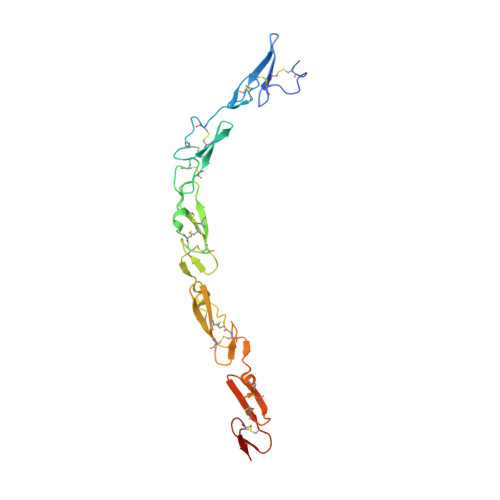
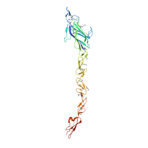
Notch-Jagged complex structure implicates a catch bond in tuning ligand sensitivity.

Luca, V.C.Kim, B.C.Ge, C.Kakuda, S.Wu, D.Roein-Peikar, M.Haltiwanger, R.S.Zhu, C.Ha, T.Garcia, K.C.

(2017) Science 355: 1320-1324

  • DOI: https://doi.org/10.1126/science.aaf9739
  • Primary Citation of Related Structures:  
    5UK5

  • PubMed Abstract: 

    Notch receptor activation initiates cell fate decisions and is distinctive in its reliance on mechanical force and protein glycosylation. The 2.5-angstrom-resolution crystal structure of the extracellular interacting region of Notch1 complexed with an engineered, high-affinity variant of Jagged1 (Jag1) reveals a binding interface that extends ~120 angstroms along five consecutive domains of each protein. O -Linked fucose modifications on Notch1 epidermal growth factor-like (EGF) domains 8 and 12 engage the EGF3 and C2 domains of Jag1, respectively, and different Notch1 domains are favored in binding to Jag1 than those that bind to the Delta-like 4 ligand. Jag1 undergoes conformational changes upon Notch binding, exhibiting catch bond behavior that prolongs interactions in the range of forces required for Notch activation. This mechanism enables cellular forces to regulate binding, discriminate among Notch ligands, and potentiate Notch signaling.


  • Organizational Affiliation

    Departments of Molecular and Cellular Physiology and Structural Biology, Stanford University School of Medicine, Stanford, CA 94305, USA.


Macromolecules
Find similar proteins by:  (by identity cutoff)  |  3D Structure
Entity ID: 1
MoleculeChains Sequence LengthOrganismDetailsImage
Neurogenic locus notch homolog protein 1205Rattus norvegicusMutation(s): 0 
Gene Names: Notch1
UniProt
Find proteins for Q07008 (Rattus norvegicus)
Explore Q07008 
Go to UniProtKB:  Q07008
Entity Groups  
Sequence Clusters30% Identity50% Identity70% Identity90% Identity95% Identity100% Identity
UniProt GroupQ07008
Sequence Annotations
Expand
  • Reference Sequence
Find similar proteins by:  (by identity cutoff)  |  3D Structure
Entity ID: 2
MoleculeChains Sequence LengthOrganismDetailsImage
Protein jagged-1318Rattus norvegicusMutation(s): 5 
Gene Names: Jag1
UniProt
Find proteins for Q63722 (Rattus norvegicus)
Explore Q63722 
Go to UniProtKB:  Q63722
Entity Groups  
Sequence Clusters30% Identity50% Identity70% Identity90% Identity95% Identity100% Identity
UniProt GroupQ63722
Sequence Annotations
Expand
  • Reference Sequence
Oligosaccharides

Help

Entity ID: 3
MoleculeChains Length2D Diagram Glycosylation3D Interactions
alpha-D-xylopyranose-(1-3)-beta-D-glucopyranose
C
2O-Glycosylation
Glycosylation Resources
GlyTouCan:  G01629YL
GlyCosmos:  G01629YL
GlyGen:  G01629YL
Small Molecules
Ligands 5 Unique
IDChains Name / Formula / InChI Key2D Diagram3D Interactions
NAG
Query on NAG

Download Ideal Coordinates CCD File 
J [auth A],
L [auth B]
2-acetamido-2-deoxy-beta-D-glucopyranose
C8 H15 N O6
OVRNDRQMDRJTHS-FMDGEEDCSA-N
MES
Query on MES

Download Ideal Coordinates CCD File 
N [auth B]2-(N-MORPHOLINO)-ETHANESULFONIC ACID
C6 H13 N O4 S
SXGZJKUKBWWHRA-UHFFFAOYSA-N
BGC
Query on BGC

Download Ideal Coordinates CCD File 
H [auth A],
I [auth A]
beta-D-glucopyranose
C6 H12 O6
WQZGKKKJIJFFOK-VFUOTHLCSA-N
FUC
Query on FUC

Download Ideal Coordinates CCD File 
G [auth A],
K [auth A],
M [auth B]
alpha-L-fucopyranose
C6 H12 O5
SHZGCJCMOBCMKK-SXUWKVJYSA-N
CA
Query on CA

Download Ideal Coordinates CCD File 
D [auth A],
E [auth A],
F [auth A]
CALCIUM ION
Ca
BHPQYMZQTOCNFJ-UHFFFAOYSA-N
Modified Residues  1 Unique
IDChains TypeFormula2D DiagramParent
MLY
Query on MLY
B
L-PEPTIDE LINKINGC8 H18 N2 O2LYS
Experimental Data & Validation

Experimental Data

  • Method: X-RAY DIFFRACTION
  • Resolution: 2.51 Å
  • R-Value Free: 0.263 
  • R-Value Work: 0.219 
  • R-Value Observed: 0.221 
  • Space Group: I 21 21 21
Unit Cell:
Length ( Å )Angle ( ˚ )
a = 68.457α = 90
b = 127.995β = 90
c = 154.288γ = 90
Software Package:
Software NamePurpose
PHENIXrefinement
HKL-2000data scaling
PDB_EXTRACTdata extraction
HKL-2000data reduction
PHASERphasing
DENZOdata reduction
SCALEPACKdata scaling

Structure Validation

View Full Validation Report


Ligand Structure Quality Assessment 


Entry History & Funding Information

Deposition Data


Funding OrganizationLocationGrant Number
National Institutes of Health/National Institute of General Medical Sciences (NIH/NIGMS)United StatesNIH-1R01-GM097015
Howard Hughes Medical Institute (HHMI)United States--
Ludwig Cancer FoundationUnited States--

Revision History  (Full details and data files)

  • Version 1.0: 2017-03-08
    Type: Initial release
  • Version 1.1: 2017-03-15
    Changes: Database references
  • Version 1.2: 2017-04-12
    Changes: Database references
  • Version 1.3: 2017-09-20
    Changes: Author supporting evidence, Refinement description
  • Version 1.4: 2018-01-24
    Changes: Structure summary
  • Version 1.5: 2019-11-20
    Changes: Author supporting evidence, Data collection
  • Version 2.0: 2020-07-29
    Type: Remediation
    Reason: Carbohydrate remediation
    Changes: Atomic model, Data collection, Derived calculations, Structure summary
  • Version 2.1: 2023-10-04
    Changes: Data collection, Database references, Derived calculations, Refinement description, Structure summary