5UAE

Crystal structure of the coiled-coil domain from Listeria Innocua Phage Integrase (Trigonal Form)


Experimental Data Snapshot

  • Method: X-RAY DIFFRACTION
  • Resolution: 2.75 Å
  • R-Value Free: 0.250 
  • R-Value Work: 0.216 
  • R-Value Observed: 0.221 

wwPDB Validation   3D Report Full Report


This is version 1.4 of the entry. See complete history


Literature

Coiled-coil interactions mediate serine integrase directionality.

Gupta, K.Sharp, R.Yuan, J.B.Li, H.Van Duyne, G.D.

(2017) Nucleic Acids Res 45: 7339-7353

  • DOI: https://doi.org/10.1093/nar/gkx474
  • Primary Citation of Related Structures:  
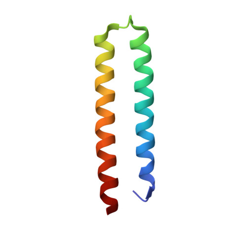
    5U96, 5UAE, 5UDO

  • PubMed Abstract: 

    Serine integrases are bacteriophage enzymes that carry out site-specific integration and excision of their viral genomes. The integration reaction is highly directional; recombination between the phage attachment site attP and the host attachment site attB to form the hybrid sites attL and attR is essentially irreversible. In a recent model, extended coiled-coil (CC) domains in the integrase subunits are proposed to interact in a way that favors the attPxattB reaction but inhibits the attLxattR reaction. Here, we show for the Listeria innocua integrase (LI Int) system that the CC domain promotes self-interaction in isolated Int and when Int is bound to attachment sites. Three independent crystal structures of the CC domain reveal the molecular nature of the CC dimer interface. Alanine substitutions of key residues in the interface support the functional significance of the structural model and indicate that the same interaction is responsible for promoting integration and for inhibiting excision. An updated model of a LI Int•attL complex that incorporates the high resolution CC dimer structure provides insights that help to explain the unusual CC dimer structure and potential sources of stability in Int•attL and Int•attR complexes. Together, the data provide a molecular basis for understanding serine integrase directionality.


  • Organizational Affiliation

    Department of Biochemistry & Biophysics, Perelman School of Medicine, University of Pennsylvania, Philadelphia, PA 10104, USA.


Macromolecules
Find similar proteins by:  (by identity cutoff)  |  3D Structure
Entity ID: 1
MoleculeChains Sequence LengthOrganismDetailsImage
Putative integrase
A, B, C, D
62Listeria innocuaMutation(s): 0 
Gene Names: int
UniProt
Find proteins for Q928V6 (Listeria innocua serovar 6a (strain ATCC BAA-680 / CLIP 11262))
Explore Q928V6 
Go to UniProtKB:  Q928V6
Entity Groups  
Sequence Clusters30% Identity50% Identity70% Identity90% Identity95% Identity100% Identity
UniProt GroupQ928V6
Sequence Annotations
Expand
  • Reference Sequence
Experimental Data & Validation

Experimental Data

  • Method: X-RAY DIFFRACTION
  • Resolution: 2.75 Å
  • R-Value Free: 0.250 
  • R-Value Work: 0.216 
  • R-Value Observed: 0.221 
  • Space Group: P 31
Unit Cell:
Length ( Å )Angle ( ˚ )
a = 75.432α = 90
b = 75.432β = 90
c = 103.061γ = 120
Software Package:
Software NamePurpose
PHENIXrefinement
HKL-3000data reduction
HKL-3000data scaling
PHENIXphasing

Structure Validation

View Full Validation Report


Entry History & Funding Information

Deposition Data


Funding OrganizationLocationGrant Number
National Institutes of Health/National Institute of General Medical Sciences (NIH/NIGMS)United States5 R01 GM108751 03

Revision History  (Full details and data files)

  • Version 1.0: 2017-05-24
    Type: Initial release
  • Version 1.1: 2017-09-27
    Changes: Author supporting evidence
  • Version 1.2: 2018-12-19
    Changes: Data collection, Database references
  • Version 1.3: 2020-01-01
    Changes: Author supporting evidence
  • Version 1.4: 2023-10-04
    Changes: Data collection, Database references, Refinement description