5U8Z

Structure of Fe-CAO1 in complex with beta-fluororesveratrol

  • Classification: OXIDOREDUCTASE
  • Organism(s): Neurospora crassa OR74A
  • Expression System: Escherichia coli
  • Mutation(s): No 

  • Deposited: 2016-12-15 Released: 2017-05-31 
  • Deposition Author(s): Sui, X., Palczewski, K., Kiser, P.D.
  • Funding Organization(s): National Institutes of Health/National Eye Institute (NIH/NEI), National Institutes of Health/National Cancer Institute (NIH/NCI), National Institutes of Health/National Institute of General Medical Sciences (NIH/NIGMS), Burroughs Wellcome Fund, Dept of Veterans Affairs

Experimental Data Snapshot

  • Method: X-RAY DIFFRACTION
  • Resolution: 1.90 Å
  • R-Value Free: 0.213 
  • R-Value Work: 0.182 
  • R-Value Observed: 0.183 

wwPDB Validation   3D Report Full Report


Ligand Structure Quality Assessment 


This is version 1.5 of the entry. See complete history


Literature

Structure and Spectroscopy of Alkene-Cleaving Dioxygenases Containing an Atypically Coordinated Non-Heme Iron Center.

Sui, X.Weitz, A.C.Farquhar, E.R.Badiee, M.Banerjee, S.von Lintig, J.Tochtrop, G.P.Palczewski, K.Hendrich, M.P.Kiser, P.D.

(2017) Biochemistry 56: 2836-2852

  • DOI: https://doi.org/10.1021/acs.biochem.7b00251
  • Primary Citation of Related Structures:  
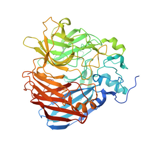
    5U8X, 5U8Y, 5U8Z, 5U90, 5U97

  • PubMed Abstract: 

    Carotenoid cleavage oxygenases (CCOs) are non-heme iron enzymes that catalyze scission of alkene groups in carotenoids and stilbenoids to form biologically important products. CCOs possess a rare four-His iron center whose resting-state structure and interaction with substrates are incompletely understood. Here, we address this knowledge gap through a comprehensive structural and spectroscopic study of three phyletically diverse CCOs. The crystal structure of a fungal stilbenoid-cleaving CCO, CAO1, reveals strong similarity between its iron center and those of carotenoid-cleaving CCOs, but with a markedly different substrate-binding cleft. These enzymes all possess a five-coordinate high-spin Fe(II) center with resting-state Fe-His bond lengths of ∼2.15 Å. This ligand set generates an iron environment more electropositive than those of other non-heme iron dioxygenases as observed by Mössbauer isomer shifts. Dioxygen (O 2 ) does not coordinate iron in the absence of substrate. Substrates bind away (∼4.7 Å) from the iron and have little impact on its electronic structure, thus excluding coordination-triggered O 2 binding. However, substrate binding does perturb the spectral properties of CCO Fe-NO derivatives, indicating proximate organic substrate and O 2 -binding sites, which might influence Fe-O 2 interactions. Together, these data provide a robust description of the CCO iron center and its interactions with substrates and substrate mimetics that illuminates commonalities as well as subtle and profound structural differences within the CCO family.


  • Organizational Affiliation

    Department of Pharmacology, School of Medicine, Case Western Reserve University , 10900 Euclid Avenue, Cleveland, Ohio 44106, United States.


Macromolecules
Find similar proteins by:  (by identity cutoff)  |  3D Structure
Entity ID: 1
MoleculeChains Sequence LengthOrganismDetailsImage
Carotenoid oxygenase 1
A, B, C, D
526Neurospora crassa OR74AMutation(s): 0 
Gene Names: cao-1NCU07008
UniProt
Find proteins for Q7S860 (Neurospora crassa (strain ATCC 24698 / 74-OR23-1A / CBS 708.71 / DSM 1257 / FGSC 987))
Explore Q7S860 
Go to UniProtKB:  Q7S860
Entity Groups  
Sequence Clusters30% Identity50% Identity70% Identity90% Identity95% Identity100% Identity
UniProt GroupQ7S860
Sequence Annotations
Expand
  • Reference Sequence
Small Molecules
Ligands 4 Unique
IDChains Name / Formula / InChI Key2D Diagram3D Interactions
83D
Query on 83D

Download Ideal Coordinates CCD File 
F [auth A],
J [auth B],
M [auth C],
P [auth D]
5-[(Z)-2-fluoro-2-(4-hydroxyphenyl)ethenyl]benzene-1,3-diol
C14 H11 F O3
PNBPFKSSWYNPPD-AUWJEWJLSA-N
DMS
Query on DMS

Download Ideal Coordinates CCD File 
H [auth A],
R [auth D]
DIMETHYL SULFOXIDE
C2 H6 O S
IAZDPXIOMUYVGZ-UHFFFAOYSA-N
FE2
Query on FE2

Download Ideal Coordinates CCD File 
E [auth A],
I [auth B],
L [auth C],
O [auth D]
FE (II) ION
Fe
CWYNVVGOOAEACU-UHFFFAOYSA-N
CL
Query on CL

Download Ideal Coordinates CCD File 
G [auth A],
K [auth B],
N [auth C],
Q [auth D]
CHLORIDE ION
Cl
VEXZGXHMUGYJMC-UHFFFAOYSA-M
Experimental Data & Validation

Experimental Data

  • Method: X-RAY DIFFRACTION
  • Resolution: 1.90 Å
  • R-Value Free: 0.213 
  • R-Value Work: 0.182 
  • R-Value Observed: 0.183 
  • Space Group: P 32 2 1
Unit Cell:
Length ( Å )Angle ( ˚ )
a = 101.037α = 90
b = 101.037β = 90
c = 448.622γ = 120
Software Package:
Software NamePurpose
REFMACrefinement
XDSdata reduction
XDSdata scaling

Structure Validation

View Full Validation Report


Ligand Structure Quality Assessment 


Entry History & Funding Information

Deposition Data


Funding OrganizationLocationGrant Number
National Institutes of Health/National Eye Institute (NIH/NEI)United StatesEY009339
National Institutes of Health/National Eye Institute (NIH/NEI)United StatesEY020551
National Institutes of Health/National Cancer Institute (NIH/NCI)CA157735
National Institutes of Health/National Institute of General Medical Sciences (NIH/NIGMS)GM77387
Burroughs Wellcome FundUnited States1015187
Dept of Veterans AffairsUnited StatesBX002683

Revision History  (Full details and data files)

  • Version 1.0: 2017-05-31
    Type: Initial release
  • Version 1.1: 2017-06-21
    Changes: Database references, Structure summary
  • Version 1.2: 2017-09-13
    Changes: Author supporting evidence
  • Version 1.3: 2019-12-11
    Changes: Author supporting evidence
  • Version 1.4: 2022-04-13
    Changes: Author supporting evidence, Database references, Derived calculations
  • Version 1.5: 2023-10-04
    Changes: Data collection, Refinement description