5U8V

Dihydrolipoamide dehydrogenase (LpdG) from Pseudomonas aeruginosa bound to NAD+


Experimental Data Snapshot

  • Method: X-RAY DIFFRACTION
  • Resolution: 1.45 Å
  • R-Value Free: 0.174 
  • R-Value Work: 0.152 
  • R-Value Observed: 0.152 

wwPDB Validation   3D Report Full Report


Ligand Structure Quality Assessment 


This is version 1.3 of the entry. See complete history


Literature

The Pyruvate and alpha-Ketoglutarate Dehydrogenase Complexes of Pseudomonas aeruginosa Catalyze Pyocyanin and Phenazine-1-carboxylic Acid Reduction via the Subunit Dihydrolipoamide Dehydrogenase.

Glasser, N.R.Wang, B.X.Hoy, J.A.Newman, D.K.

(2017) J Biol Chem 292: 5593-5607

  • DOI: https://doi.org/10.1074/jbc.M116.772848
  • Primary Citation of Related Structures:  
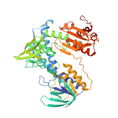
    5U8U, 5U8V, 5U8W

  • PubMed Abstract: 

    Phenazines are a class of redox-active molecules produced by diverse bacteria and archaea. Many of the biological functions of phenazines, such as mediating signaling, iron acquisition, and redox homeostasis, derive from their redox activity. Although prior studies have focused on extracellular phenazine oxidation by oxygen and iron, here we report a search for reductants and catalysts of intracellular phenazine reduction in Pseudomonas aeruginosa Enzymatic assays in cell-free lysate, together with crude fractionation and chemical inhibition, indicate that P. aeruginosa contains multiple enzymes that catalyze the reduction of the endogenous phenazines pyocyanin and phenazine-1-carboxylic acid in both cytosolic and membrane fractions. We used chemical inhibitors to target general enzyme classes and found that an inhibitor of flavoproteins and heme-containing proteins, diphenyleneiodonium, effectively inhibited phenazine reduction in vitro , suggesting that most phenazine reduction derives from these enzymes. Using natively purified proteins, we demonstrate that the pyruvate and α-ketoglutarate dehydrogenase complexes directly catalyze phenazine reduction with pyruvate or α-ketoglutarate as electron donors. Both complexes transfer electrons to phenazines through the common subunit dihydrolipoamide dehydrogenase, a flavoprotein encoded by the gene lpdG Although we were unable to co-crystallize LpdG with an endogenous phenazine, we report its X-ray crystal structure in the apo-form (refined to 1.35 Å), bound to NAD + (1.45 Å), and bound to NADH (1.79 Å). In contrast to the notion that phenazines support intracellular redox homeostasis by oxidizing NADH, our work suggests that phenazines may substitute for NAD + in LpdG and other enzymes, achieving the same end by a different mechanism.


  • Organizational Affiliation

    From the Divisions of Biology and Biological Engineering and.


Macromolecules
Find similar proteins by:  (by identity cutoff)  |  3D Structure
Entity ID: 1
MoleculeChains Sequence LengthOrganismDetailsImage
Dihydrolipoyl dehydrogenase
A, B, C, D
481Pseudomonas aeruginosa UCBPP-PA14Mutation(s): 0 
Gene Names: lpdGPA14_43970
EC: 1.8.1.4
UniProt
Find proteins for A0A0H2Z9F5 (Pseudomonas aeruginosa (strain UCBPP-PA14))
Explore A0A0H2Z9F5 
Go to UniProtKB:  A0A0H2Z9F5
Entity Groups  
Sequence Clusters30% Identity50% Identity70% Identity90% Identity95% Identity100% Identity
UniProt GroupA0A0H2Z9F5
Sequence Annotations
Expand
  • Reference Sequence
Small Molecules
Ligands 3 Unique
IDChains Name / Formula / InChI Key2D Diagram3D Interactions
FAD
Query on FAD

Download Ideal Coordinates CCD File 
E [auth A],
JA [auth D],
P [auth B],
Y [auth C]
FLAVIN-ADENINE DINUCLEOTIDE
C27 H33 N9 O15 P2
VWWQXMAJTJZDQX-UYBVJOGSSA-N
NAD
Query on NAD

Download Ideal Coordinates CCD File 
F [auth A],
KA [auth D],
Q [auth B],
Z [auth C]
NICOTINAMIDE-ADENINE-DINUCLEOTIDE
C21 H27 N7 O14 P2
BAWFJGJZGIEFAR-NNYOXOHSSA-N
DMS
Query on DMS

Download Ideal Coordinates CCD File 
AA [auth C]
BA [auth C]
CA [auth C]
DA [auth C]
EA [auth C]
AA [auth C],
BA [auth C],
CA [auth C],
DA [auth C],
EA [auth C],
FA [auth C],
G [auth A],
GA [auth C],
H [auth A],
HA [auth C],
I [auth A],
IA [auth C],
J [auth A],
K [auth A],
L [auth A],
LA [auth D],
M [auth A],
MA [auth D],
N [auth A],
NA [auth D],
O [auth B],
OA [auth D],
PA [auth D],
QA [auth D],
R [auth B],
RA [auth D],
S [auth B],
T [auth B],
U [auth B],
V [auth B],
W [auth B],
X [auth B]
DIMETHYL SULFOXIDE
C2 H6 O S
IAZDPXIOMUYVGZ-UHFFFAOYSA-N
Binding Affinity Annotations 
IDSourceBinding Affinity
FAD Binding MOAD:  5U8V Kd: 8.40e+7 (nM) from 1 assay(s)
Experimental Data & Validation

Experimental Data

  • Method: X-RAY DIFFRACTION
  • Resolution: 1.45 Å
  • R-Value Free: 0.174 
  • R-Value Work: 0.152 
  • R-Value Observed: 0.152 
  • Space Group: P 1 21 1
Unit Cell:
Length ( Å )Angle ( ˚ )
a = 65.61α = 90
b = 116.69β = 94.87
c = 136.86γ = 90
Software Package:
Software NamePurpose
PHENIXrefinement
XDSdata reduction
XSCALEdata scaling
PHASERphasing

Structure Validation

View Full Validation Report


Ligand Structure Quality Assessment 


Entry History 

Deposition Data

Revision History  (Full details and data files)

  • Version 1.0: 2017-02-15
    Type: Initial release
  • Version 1.1: 2017-02-22
    Changes: Database references
  • Version 1.2: 2017-04-12
    Changes: Database references
  • Version 1.3: 2023-10-04
    Changes: Data collection, Database references, Refinement description