5U6F

Bacterial adhesin from Mobiluncus mulieris containing intramolecular disulfide, isopeptide, and ester bond cross-links (space group P21)


Experimental Data Snapshot

  • Method: X-RAY DIFFRACTION
  • Resolution: 1.50 Å
  • R-Value Free: 0.267 
  • R-Value Work: 0.234 
  • R-Value Observed: 0.235 

wwPDB Validation   3D Report Full Report


This is version 1.1 of the entry. See complete history


Literature

Domain structure and cross-linking in a giant adhesin from the Mobiluncus mulieris bacterium.

Young, P.G.Paynter, J.M.Wardega, J.K.Middleditch, M.J.Payne, L.S.Baker, E.N.Squire, C.J.

(2023) Acta Crystallogr D Struct Biol 79: 971-979

  • DOI: https://doi.org/10.1107/S2059798323007507
  • Primary Citation of Related Structures:  
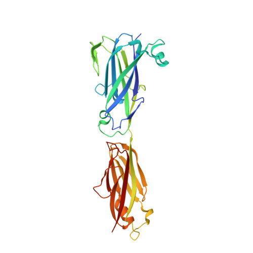
    5U5O, 5U6F

  • PubMed Abstract: 

    Cell-surface proteins known as adhesins enable bacteria to colonize particular environments, and in Gram-positive bacteria often contain autocatalytically formed covalent intramolecular cross-links. While investigating the prevalence of such cross-links, a remarkable example was discovered in Mobiluncus mulieris, a pathogen associated with bacterial vaginosis. This organism encodes a putative adhesin of 7651 residues. Crystallography and mass spectrometry of two selected domains, and AlphaFold structure prediction of the remainder of the protein, were used to show that this adhesin belongs to the family of thioester, isopeptide and ester-bond-containing proteins (TIE proteins). It has an N-terminal domain homologous to thioester adhesion domains, followed by 51 immunoglobulin (Ig)-like domains containing ester- or isopeptide-bond cross-links. The energetic cost to the M. mulieris bacterium in retaining such a large adhesin as a single gene or protein construct suggests a critical role in pathogenicity and/or persistence.


  • Organizational Affiliation

    School of Biological Sciences, The University of Auckland, Private Bag 92019, Auckland 1010, New Zealand.


Macromolecules
Find similar proteins by:  (by identity cutoff)  |  3D Structure
Entity ID: 1
MoleculeChains Sequence LengthOrganismDetailsImage
LPXTG-motif cell wall anchor domain protein292Mobiluncus mulierisMutation(s): 0 
UniProt
Find proteins for E0QN07 (Mobiluncus mulieris ATCC 35239)
Explore E0QN07 
Go to UniProtKB:  E0QN07
Entity Groups  
Sequence Clusters30% Identity50% Identity70% Identity90% Identity95% Identity100% Identity
UniProt GroupE0QN07
Sequence Annotations
Expand
  • Reference Sequence
Experimental Data & Validation

Experimental Data

  • Method: X-RAY DIFFRACTION
  • Resolution: 1.50 Å
  • R-Value Free: 0.267 
  • R-Value Work: 0.234 
  • R-Value Observed: 0.235 
  • Space Group: P 1 21 1
Unit Cell:
Length ( Å )Angle ( ˚ )
a = 34.548α = 90
b = 51.604β = 101.6
c = 81.206γ = 90
Software Package:
Software NamePurpose
REFMACrefinement
XDSdata reduction
Aimlessdata scaling
PHASERphasing

Structure Validation

View Full Validation Report


Entry History 

Deposition Data

Revision History  (Full details and data files)

  • Version 1.0: 2018-06-13
    Type: Initial release
  • Version 1.1: 2024-04-03
    Changes: Data collection, Database references, Refinement description