5TXR

Structure of ALAS from S. cerevisiae non-covalently bound to PLP cofactor

  • Classification: TRANSFERASE
  • Organism(s): Saccharomyces cerevisiae S288C
  • Expression System: Escherichia coli
  • Mutation(s): No 

  • Deposited: 2016-11-17 Released: 2018-03-28 
  • Deposition Author(s): Brown, B.L., Grant, R.A., Kardon, J.R., Sauer, R.T., Baker, T.A.
  • Funding Organization(s): Howard Hughes Medical Institute (HHMI), Burroughs Wellcome Fund, Jane Coffin Childs Memorial Fund for Medical Research, National Institutes of Health/National Institute Of Allergy and Infectious Diseases (NIH/NIAID), National Institutes of Health/National Institute of Diabetes and Digestive and Kidney Disease (NIH/NIDDK)

Experimental Data Snapshot

  • Method: X-RAY DIFFRACTION
  • Resolution: 1.90 Å
  • R-Value Free: 0.172 
  • R-Value Work: 0.142 
  • R-Value Observed: 0.142 

wwPDB Validation   3D Report Full Report


Ligand Structure Quality Assessment 


This is version 1.5 of the entry. See complete history


Literature

Structure of the Mitochondrial Aminolevulinic Acid Synthase, a Key Heme Biosynthetic Enzyme.

Brown, B.L.Kardon, J.R.Sauer, R.T.Baker, T.A.

(2018) Structure 26: 580-589.e4

  • DOI: https://doi.org/10.1016/j.str.2018.02.012
  • Primary Citation of Related Structures:  
    5TXR, 5TXT

  • PubMed Abstract: 

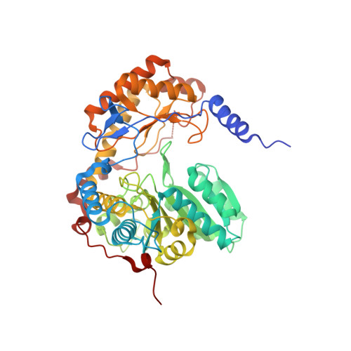
    5-Aminolevulinic acid synthase (ALAS) catalyzes the first step in heme biosynthesis. We present the crystal structure of a eukaryotic ALAS from Saccharomyces cerevisiae. In this homodimeric structure, one ALAS subunit contains covalently bound cofactor, pyridoxal 5'-phosphate (PLP), whereas the second is PLP free. Comparison between the subunits reveals PLP-coupled reordering of the active site and of additional regions to achieve the active conformation of the enzyme. The eukaryotic C-terminal extension, a region altered in multiple human disease alleles, wraps around the dimer and contacts active-site-proximal residues. Mutational analysis demonstrates that this C-terminal region that engages the active site is important for ALAS activity. Our discovery of structural elements that change conformation upon PLP binding and of direct contact between the C-terminal extension and the active site thus provides a structural basis for investigation of disruptions in the first step of heme biosynthesis and resulting human disorders.


  • Organizational Affiliation

    Department of Biology, Massachusetts Institute of Technology, Cambridge, MA 02139, USA.


Macromolecules
Find similar proteins by:  (by identity cutoff)  |  3D Structure
Entity ID: 1
MoleculeChains Sequence LengthOrganismDetailsImage
5-aminolevulinate synthase, mitochondrialA [auth B],
B [auth A]
491Saccharomyces cerevisiae S288CMutation(s): 0 
Gene Names: HEM1CYD1YDR232WYD9934.16
EC: 2.3.1.37
UniProt
Find proteins for P09950 (Saccharomyces cerevisiae (strain ATCC 204508 / S288c))
Explore P09950 
Go to UniProtKB:  P09950
Entity Groups  
Sequence Clusters30% Identity50% Identity70% Identity90% Identity95% Identity100% Identity
UniProt GroupP09950
Sequence Annotations
Expand
  • Reference Sequence
Small Molecules
Ligands 4 Unique
IDChains Name / Formula / InChI Key2D Diagram3D Interactions
PLP
Query on PLP

Download Ideal Coordinates CCD File 
G [auth B],
P [auth A]
PYRIDOXAL-5'-PHOSPHATE
C8 H10 N O6 P
NGVDGCNFYWLIFO-UHFFFAOYSA-N
GOL
Query on GOL

Download Ideal Coordinates CCD File 
H [auth A]GLYCEROL
C3 H8 O3
PEDCQBHIVMGVHV-UHFFFAOYSA-N
FMT
Query on FMT

Download Ideal Coordinates CCD File 
C [auth B]
D [auth B]
E [auth B]
F [auth B]
J [auth A]
C [auth B],
D [auth B],
E [auth B],
F [auth B],
J [auth A],
K [auth A],
L [auth A],
M [auth A],
N [auth A],
O [auth A]
FORMIC ACID
C H2 O2
BDAGIHXWWSANSR-UHFFFAOYSA-N
K
Query on K

Download Ideal Coordinates CCD File 
I [auth A]POTASSIUM ION
K
NPYPAHLBTDXSSS-UHFFFAOYSA-N
Experimental Data & Validation

Experimental Data

  • Method: X-RAY DIFFRACTION
  • Resolution: 1.90 Å
  • R-Value Free: 0.172 
  • R-Value Work: 0.142 
  • R-Value Observed: 0.142 
  • Space Group: P 1 21 1
Unit Cell:
Length ( Å )Angle ( ˚ )
a = 78.613α = 90
b = 64.878β = 95.86
c = 95.693γ = 90
Software Package:
Software NamePurpose
PHENIXrefinement
HKL-2000data reduction
HKL-2000data scaling
PHENIXphasing

Structure Validation

View Full Validation Report


Ligand Structure Quality Assessment 


Entry History & Funding Information

Deposition Data


Funding OrganizationLocationGrant Number
Howard Hughes Medical Institute (HHMI)United States--
Burroughs Wellcome FundUnited States1015092
Jane Coffin Childs Memorial Fund for Medical ResearchUnited States61-1529
National Institutes of Health/National Institute Of Allergy and Infectious Diseases (NIH/NIAID)United StatesAI-16892
National Institutes of Health/National Institute of Diabetes and Digestive and Kidney Disease (NIH/NIDDK)United StatesF32DK095726

Revision History  (Full details and data files)

  • Version 1.0: 2018-03-28
    Type: Initial release
  • Version 1.1: 2018-04-11
    Changes: Data collection, Database references
  • Version 1.2: 2018-04-18
    Changes: Data collection, Database references
  • Version 1.3: 2019-11-20
    Changes: Author supporting evidence
  • Version 1.4: 2024-03-06
    Changes: Data collection, Database references
  • Version 1.5: 2024-04-03
    Changes: Refinement description