5TEJ

Structure of 4-Hydroxy-tetrahydrodipicolinate Reductase from Vibrio vulnificus with 2,5 Furan Dicarboxylic and NADH


Experimental Data Snapshot

  • Method: X-RAY DIFFRACTION
  • Resolution: 2.50 Å
  • R-Value Free: 0.212 
  • R-Value Work: 0.182 
  • R-Value Observed: 0.183 

wwPDB Validation   3D Report Full Report


Ligand Structure Quality Assessment 


This is version 1.2 of the entry. See complete history


Literature

Comparative structural and mechanistic studies of 4-hydroxy-tetrahydrodipicolinate reductases from Mycobacterium tuberculosis and Vibrio vulnificus.

Pote, S.Kachhap, S.Mank, N.J.Daneshian, L.Klapper, V.Pye, S.Arnette, A.K.Shimizu, L.S.Borowski, T.Chruszcz, M.

(2021) Biochim Biophys Acta Gen Subj 1865: 129750-129750

  • DOI: https://doi.org/10.1016/j.bbagen.2020.129750
  • Primary Citation of Related Structures:  
    5TEJ, 5TEK, 5TEM, 5TEN, 5TJY, 5TJZ, 5UGV, 5US6

  • PubMed Abstract: 

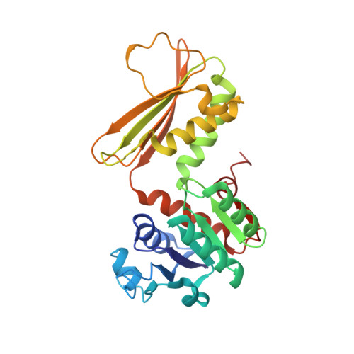
    The products of the lysine biosynthesis pathway, meso-diaminopimelate and lysine, are essential for bacterial survival. This paper focuses on the structural and mechanistic characterization of 4-hydroxy-tetrahydrodipicolinate reductase (DapB), which is one of the enzymes from the lysine biosynthesis pathway. DapB catalyzes the conversion of (2S, 4S)-4-hydroxy-2,3,4,5-tetrahydrodipicolinate (HTPA) to 2,3,4,5-tetrahydrodipicolinate in an NADH/NADPH dependent reaction. Genes coding for DapBs were identified as essential for many pathogenic bacteria, and therefore DapB is an interesting new target for the development of antibiotics.


  • Organizational Affiliation

    Department of Chemistry and Biochemistry, University of South Carolina, Columbia, SC 29208, USA.


Macromolecules
Find similar proteins by:  (by identity cutoff)  |  3D Structure
Entity ID: 1
MoleculeChains Sequence LengthOrganismDetailsImage
4-hydroxy-tetrahydrodipicolinate reductase
A, B, C, D
270Vibrio vulnificus CMCP6Mutation(s): 0 
Gene Names: dapBVV1_0567
EC: 1.17.1.8
UniProt
Find proteins for Q8DEM0 (Vibrio vulnificus (strain CMCP6))
Explore Q8DEM0 
Go to UniProtKB:  Q8DEM0
Entity Groups  
Sequence Clusters30% Identity50% Identity70% Identity90% Identity95% Identity100% Identity
UniProt GroupQ8DEM0
Sequence Annotations
Expand
  • Reference Sequence
Small Molecules
Ligands 5 Unique
IDChains Name / Formula / InChI Key2D Diagram3D Interactions
NAD
Query on NAD

Download Ideal Coordinates CCD File 
E [auth A],
G [auth B],
K [auth C],
N [auth D]
NICOTINAMIDE-ADENINE-DINUCLEOTIDE
C21 H27 N7 O14 P2
BAWFJGJZGIEFAR-NNYOXOHSSA-N
PG4
Query on PG4

Download Ideal Coordinates CCD File 
F [auth A]TETRAETHYLENE GLYCOL
C8 H18 O5
UWHCKJMYHZGTIT-UHFFFAOYSA-N
7FN
Query on 7FN

Download Ideal Coordinates CCD File 
H [auth B],
L [auth C],
O [auth D]
2,5 Furan Dicarboxylic Acid
C6 H4 O5
CHTHALBTIRVDBM-UHFFFAOYSA-N
PGE
Query on PGE

Download Ideal Coordinates CCD File 
J [auth B]TRIETHYLENE GLYCOL
C6 H14 O4
ZIBGPFATKBEMQZ-UHFFFAOYSA-N
NA
Query on NA

Download Ideal Coordinates CCD File 
I [auth B],
M [auth C],
P [auth D],
Q [auth D]
SODIUM ION
Na
FKNQFGJONOIPTF-UHFFFAOYSA-N
Experimental Data & Validation

Experimental Data

  • Method: X-RAY DIFFRACTION
  • Resolution: 2.50 Å
  • R-Value Free: 0.212 
  • R-Value Work: 0.182 
  • R-Value Observed: 0.183 
  • Space Group: C 1 2 1
Unit Cell:
Length ( Å )Angle ( ˚ )
a = 127.059α = 90
b = 91.039β = 120.07
c = 125.922γ = 90
Software Package:
Software NamePurpose
REFMACrefinement
HKL-3000data reduction
HKL-3000data scaling
HKL-3000phasing

Structure Validation

View Full Validation Report


Ligand Structure Quality Assessment 


Entry History 

Deposition Data

Revision History  (Full details and data files)

  • Version 1.0: 2017-10-18
    Type: Initial release
  • Version 1.1: 2021-06-30
    Changes: Database references, Derived calculations
  • Version 1.2: 2023-10-04
    Changes: Data collection, Database references, Refinement description