5TDD

Human UBR-box from UBR2 in complex with HIFS peptide


Experimental Data Snapshot

  • Method: X-RAY DIFFRACTION
  • Resolution: 1.55 Å
  • R-Value Free: 0.152 
  • R-Value Work: 0.106 
  • R-Value Observed: 0.111 

wwPDB Validation   3D Report Full Report


This is version 1.5 of the entry. See complete history


Literature

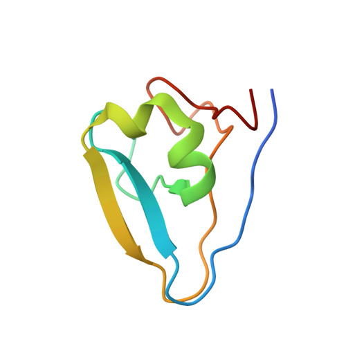
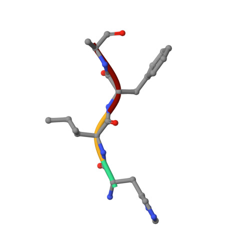
Bound Waters Mediate Binding of Diverse Substrates to a Ubiquitin Ligase.

Munoz-Escobar, J.Matta-Camacho, E.Cho, C.Kozlov, G.Gehring, K.

(2017) Structure 25: 719-729.e3

  • DOI: https://doi.org/10.1016/j.str.2017.03.004
  • Primary Citation of Related Structures:  
    5TDA, 5TDB, 5TDC, 5TDD, 5UM3

  • PubMed Abstract: 

    The N-end rule pathway controls the half-life of proteins based on their N-terminal residue. Positively charged type 1 N-degrons are recognized by a negatively charged pocket on the Zn finger named the UBR box. Here, we show that the UBR box is rigid, but bound water molecules in the pocket provide the structural plasticity required to bind different positively charged amino acids. Ultra-high-resolution crystal structures of arginine, histidine, and methylated arginine reveal that water molecules mediate the binding of N-degron peptides. Using a high-throughput binding assay and isothermal titration calorimetry, we demonstrate that the UBR box is able to bind methylated arginine and lysine peptides with high affinity and measure the preference for hydrophobic residues in the second position in the N-degron peptide. Finally, we show that the V122L mutation present in Johanson-Blizzard syndrome patients changes the specificity for the second position due to occlusion of the secondary pocket.


  • Organizational Affiliation

    Department of Biochemistry, Groupe de Recherche Axé sur la Structure des Protéines, McGill University, Montreal, QC H3G 0B1, Canada.


Macromolecules
Find similar proteins by:  (by identity cutoff)  |  3D Structure
Entity ID: 1
MoleculeChains Sequence LengthOrganismDetailsImage
E3 ubiquitin-protein ligase UBR276Homo sapiensMutation(s): 0 
Gene Names: UBR2C6orf133KIAA0349
EC: 6.3.2
UniProt & NIH Common Fund Data Resources
Find proteins for Q8IWV8 (Homo sapiens)
Explore Q8IWV8 
Go to UniProtKB:  Q8IWV8
PHAROS:  Q8IWV8
GTEx:  ENSG00000024048 
Entity Groups  
Sequence Clusters30% Identity50% Identity70% Identity90% Identity95% Identity100% Identity
UniProt GroupQ8IWV8
Sequence Annotations
Expand
  • Reference Sequence

Find similar proteins by:  Sequence   |   3D Structure  

Entity ID: 2
MoleculeChains Sequence LengthOrganismDetailsImage
HIS-ILE-PHE-SER peptide4Homo sapiensMutation(s): 0 
Sequence Annotations
Expand
  • Reference Sequence
Experimental Data & Validation

Experimental Data

  • Method: X-RAY DIFFRACTION
  • Resolution: 1.55 Å
  • R-Value Free: 0.152 
  • R-Value Work: 0.106 
  • R-Value Observed: 0.111 
  • Space Group: P 41
Unit Cell:
Length ( Å )Angle ( ˚ )
a = 29.313α = 90
b = 29.313β = 90
c = 74.151γ = 90
Software Package:
Software NamePurpose
PHENIXrefinement
HKL-2000data reduction
HKL-2000data scaling
PHASERphasing

Structure Validation

View Full Validation Report


Entry History & Funding Information

Deposition Data


Funding OrganizationLocationGrant Number
Canadian Institutes of Health Research (CIHR)Canada--

Revision History  (Full details and data files)

  • Version 1.0: 2017-03-22
    Type: Initial release
  • Version 1.1: 2017-04-26
    Changes: Database references
  • Version 1.2: 2017-05-10
    Changes: Database references
  • Version 1.3: 2017-09-20
    Changes: Author supporting evidence
  • Version 1.4: 2020-01-08
    Changes: Author supporting evidence
  • Version 1.5: 2023-10-04
    Changes: Data collection, Database references, Derived calculations, Refinement description