5SZ8

Truncated hemolysin A Q125A/Y134A from P. mirabilis at 1.8 Angstroms resolution crystallized in a high salt condition


Experimental Data Snapshot

  • Method: X-RAY DIFFRACTION
  • Resolution: 1.83 Å
  • R-Value Free: 0.206 
  • R-Value Work: 0.178 
  • R-Value Observed: 0.179 

wwPDB Validation   3D Report Full Report


This is version 1.4 of the entry. See complete history


Literature

Proteolysis of truncated hemolysin A yields a stable dimerization interface.

Novak, W.R.Bhattacharyya, B.Grilley, D.P.Weaver, T.M.

(2017) Acta Crystallogr F Struct Biol Commun 73: 138-145

  • DOI: https://doi.org/10.1107/S2053230X17002102
  • Primary Citation of Related Structures:  
    4W8Q, 5KEH, 5KF3, 5KKD, 5SZ8

  • PubMed Abstract: 

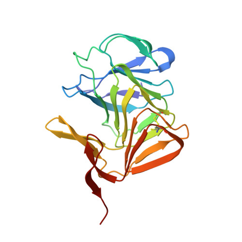
    Wild-type and variant forms of HpmA265 (truncated hemolysin A) from Proteus mirabilis reveal a right-handed, parallel β-helix capped and flanked by segments of antiparallel β-strands. The low-salt crystal structures form a dimeric structure via the implementation of on-edge main-chain hydrogen bonds donated by residues 243-263 of adjacent monomers. Surprisingly, in the high-salt structures of two variants, Y134A and Q125A-Y134A, a new dimeric interface is formed via main-chain hydrogen bonds donated by residues 203-215 of adjacent monomers, and a previously unobserved tetramer is formed. In addition, an eight-stranded antiparallel β-sheet is formed from the flap regions of crystallographically related monomers in the high-salt structures. This new interface is possible owing to additional proteolysis of these variants after Tyr240. The interface formed in the high-salt crystal forms of hemolysin A variants may mimic the on-edge β-strand positioning used in template-assisted hemolytic activity.


  • Organizational Affiliation

    Department of Chemistry, Wabash College, 301 West Wabash Avenue, Crawfordsville, IN 47933, USA.


Macromolecules
Find similar proteins by:  (by identity cutoff)  |  3D Structure
Entity ID: 1
MoleculeChains Sequence LengthOrganismDetailsImage
Hemolysin
A, B
205Proteus mirabilisMutation(s): 2 
Gene Names: hpmA
UniProt
Find proteins for P16466 (Proteus mirabilis)
Explore P16466 
Go to UniProtKB:  P16466
Entity Groups  
Sequence Clusters30% Identity50% Identity70% Identity90% Identity95% Identity100% Identity
UniProt GroupP16466
Sequence Annotations
Expand
  • Reference Sequence
Experimental Data & Validation

Experimental Data

  • Method: X-RAY DIFFRACTION
  • Resolution: 1.83 Å
  • R-Value Free: 0.206 
  • R-Value Work: 0.178 
  • R-Value Observed: 0.179 
  • Space Group: P 41 21 2
Unit Cell:
Length ( Å )Angle ( ˚ )
a = 69.231α = 90
b = 69.231β = 90
c = 234.862γ = 90
Software Package:
Software NamePurpose
PHENIXrefinement
SCALEPACKdata scaling
PHASERphasing
HKL-3000data reduction

Structure Validation

View Full Validation Report


Entry History & Funding Information

Deposition Data


Funding OrganizationLocationGrant Number
National Science Foundation (NSF, United States)United StatesMCB1050435

Revision History  (Full details and data files)

  • Version 1.0: 2017-03-22
    Type: Initial release
  • Version 1.1: 2017-05-17
    Changes: Database references
  • Version 1.2: 2017-09-20
    Changes: Author supporting evidence
  • Version 1.3: 2019-11-27
    Changes: Author supporting evidence
  • Version 1.4: 2023-10-04
    Changes: Data collection, Database references, Refinement description