5SW5

Crystal structure of native catalase-peroxidase KatG at pH7.5


Experimental Data Snapshot

  • Method: X-RAY DIFFRACTION
  • Resolution: 2.05 Å
  • R-Value Free: 0.184 
  • R-Value Work: 0.149 
  • R-Value Observed: 0.150 

wwPDB Validation   3D Report Full Report


Ligand Structure Quality Assessment 


This is version 1.4 of the entry. See complete history


Literature

A molecular switch and electronic circuit modulate catalase activity in catalase-peroxidases.

Carpena, X.Wiseman, B.Deemagarn, T.Singh, R.Switala, J.Ivancich, A.Fita, I.Loewen, P.C.

(2005) EMBO Rep 6: 1156-1162

  • DOI: https://doi.org/10.1038/sj.embor.7400550
  • Primary Citation of Related Structures:  
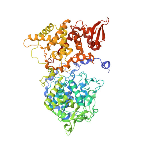
    5SW4, 5SW5, 5SW6, 5SX0

  • PubMed Abstract: 

    The catalase reaction of catalase-peroxidases involves catalase-specific features built into a peroxidase core. An arginine, 20 A from the active-site heme, acts as a molecular switch moving between two conformations, one that activates heme oxidation and one that activates oxoferryl heme reduction by H(2)O(2), facilitating the catalatic pathway in a peroxidase. The influence of the arginine is imparted to the heme through its association with or dissociation from a tyrosinate that modulates reactivity through a Met-Tyr-Trp crosslinked adduct and a pi electron interaction of the heme with the adduct Trp.


  • Organizational Affiliation

    Department of Microbiology, University of Manitoba, Winnipeg MB R3T 2N2, Canada.


Macromolecules
Find similar proteins by:  (by identity cutoff)  |  3D Structure
Entity ID: 1
MoleculeChains Sequence LengthOrganismDetailsImage
Catalase-peroxidase
A, B
728Burkholderia pseudomallei 1710bMutation(s): 0 
Gene Names: katGBURPS1710b_3366
EC: 1.11.1.21
UniProt
Find proteins for Q3JNW6 (Burkholderia pseudomallei (strain 1710b))
Explore Q3JNW6 
Go to UniProtKB:  Q3JNW6
Entity Groups  
Sequence Clusters30% Identity50% Identity70% Identity90% Identity95% Identity100% Identity
UniProt GroupQ3JNW6
Sequence Annotations
Expand
  • Reference Sequence
Small Molecules
Ligands 6 Unique
IDChains Name / Formula / InChI Key2D Diagram3D Interactions
HEM
Query on HEM

Download Ideal Coordinates CCD File 
C [auth A],
L [auth B]
PROTOPORPHYRIN IX CONTAINING FE
C34 H32 Fe N4 O4
KABFMIBPWCXCRK-RGGAHWMASA-L
TRS
Query on TRS

Download Ideal Coordinates CCD File 
I [auth A],
J [auth A],
K [auth A],
Q [auth B]
2-AMINO-2-HYDROXYMETHYL-PROPANE-1,3-DIOL
C4 H12 N O3
LENZDBCJOHFCAS-UHFFFAOYSA-O
MPD
Query on MPD

Download Ideal Coordinates CCD File 
G [auth A],
H [auth A],
P [auth B]
(4S)-2-METHYL-2,4-PENTANEDIOL
C6 H14 O2
SVTBMSDMJJWYQN-YFKPBYRVSA-N
PO4
Query on PO4

Download Ideal Coordinates CCD File 
F [auth A],
O [auth B]
PHOSPHATE ION
O4 P
NBIIXXVUZAFLBC-UHFFFAOYSA-K
OXY
Query on OXY

Download Ideal Coordinates CCD File 
E [auth A],
N [auth B]
OXYGEN MOLECULE
O2
MYMOFIZGZYHOMD-UHFFFAOYSA-N
NA
Query on NA

Download Ideal Coordinates CCD File 
D [auth A],
M [auth B]
SODIUM ION
Na
FKNQFGJONOIPTF-UHFFFAOYSA-N
Modified Residues  1 Unique
IDChains TypeFormula2D DiagramParent
TOX
Query on TOX
A, B
L-PEPTIDE LINKINGC11 H12 N2 O4TRP
Experimental Data & Validation

Experimental Data

  • Method: X-RAY DIFFRACTION
  • Resolution: 2.05 Å
  • R-Value Free: 0.184 
  • R-Value Work: 0.149 
  • R-Value Observed: 0.150 
  • Space Group: P 21 21 21
Unit Cell:
Length ( Å )Angle ( ˚ )
a = 100.343α = 90
b = 114.851β = 90
c = 174.86γ = 90
Software Package:
Software NamePurpose
REFMACrefinement
PDB_EXTRACTdata extraction
DENZOdata reduction
SCALEPACKdata scaling
AMoREphasing

Structure Validation

View Full Validation Report


Ligand Structure Quality Assessment 


Entry History & Funding Information

Deposition Data

  • Released Date: 2016-08-24 
  • Deposition Author(s): Loewen, P.C.
  • This entry supersedes: 2B2Q

Funding OrganizationLocationGrant Number
Natural Sciences and Engineering Research Council (NSERC, Canada)CanadaD9600

Revision History  (Full details and data files)

  • Version 1.0: 2016-08-24
    Type: Initial release
  • Version 1.1: 2017-09-27
    Changes: Author supporting evidence, Derived calculations, Structure summary
  • Version 1.2: 2020-01-08
    Changes: Author supporting evidence
  • Version 1.3: 2023-10-04
    Changes: Data collection, Database references, Derived calculations, Refinement description
  • Version 1.4: 2023-11-15
    Changes: Data collection