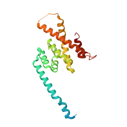
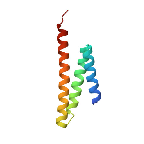
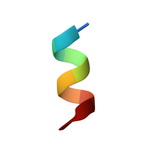
5OW5

p60p80-CAMSAP complex


Experimental Data Snapshot

  • Method: X-RAY DIFFRACTION
  • Resolution: 1.70 Å
  • R-Value Free: 0.232 
  • R-Value Work: 0.191 
  • R-Value Observed: 0.191 

wwPDB Validation   3D Report Full Report


This is version 1.1 of the entry. See complete history


Literature

Structural Basis of Formation of the Microtubule Minus-End-Regulating CAMSAP-Katanin Complex.

Jiang, K.Faltova, L.Hua, S.Capitani, G.Prota, A.E.Landgraf, C.Volkmer, R.Kammerer, R.A.Steinmetz, M.O.Akhmanova, A.

(2018) Structure 26: 375-382.e4

  • DOI: https://doi.org/10.1016/j.str.2017.12.017
  • Primary Citation of Related Structures:  
    5OW5

  • PubMed Abstract: 

    CAMSAP/Patronin family members regulate the organization and stability of microtubule minus ends in various systems ranging from mitotic spindles to differentiated epithelial cells and neurons. Mammalian CAMSAP2 and CAMSAP3 bind to growing microtubule minus ends, where they form stretches of stabilized microtubule lattice. The microtubule-severing ATPase katanin interacts with CAMSAPs and limits the length of CAMSAP-decorated microtubule stretches. Here, by using biochemical, biophysical, and structural approaches, we reveal that a short helical motif conserved in CAMSAP2 and CAMSAP3 binds to the heterodimer formed by the N- and C-terminal domains of katanin subunits p60 and p80, respectively. The identified CAMSAP-katanin binding mode is supported by mutational analysis and genome-editing experiments. It is strikingly similar to the one seen in the ASPM-katanin complex, which is responsible for microtubule minus-end regulation in mitotic spindles. Our work provides a general molecular mechanism for the cooperation of katanin with major microtubule minus-end regulators.


  • Organizational Affiliation

    Cell Biology, Department of Biology, Faculty of Science, Utrecht University, Padualaan 8, 3584 Utrecht, the Netherlands.


Macromolecules
Find similar proteins by:  (by identity cutoff)  |  3D Structure
Entity ID: 1
MoleculeChains Sequence LengthOrganismDetailsImage
Katanin p80 WD40 repeat-containing subunit B1
A, C
212Mus musculusMutation(s): 0 
Gene Names: Katnb1
UniProt & NIH Common Fund Data Resources
Find proteins for Q8BG40 (Mus musculus)
Explore Q8BG40 
Go to UniProtKB:  Q8BG40
IMPC:  MGI:1921437
Entity Groups  
Sequence Clusters30% Identity50% Identity70% Identity90% Identity95% Identity100% Identity
UniProt GroupQ8BG40
Sequence Annotations
Expand
  • Reference Sequence
Find similar proteins by:  (by identity cutoff)  |  3D Structure
Entity ID: 2
MoleculeChains Sequence LengthOrganismDetailsImage
Katanin p60 ATPase-containing subunit A1
B, D
80Mus musculusMutation(s): 0 
Gene Names: Katna1KATNA1
EC: 3.6.4.3
UniProt & NIH Common Fund Data Resources
Find proteins for E9PZI6 (Mus musculus)
Explore E9PZI6 
Go to UniProtKB:  E9PZI6
IMPC:  MGI:1344353
Entity Groups  
Sequence Clusters30% Identity50% Identity70% Identity90% Identity95% Identity100% Identity
UniProt GroupE9PZI6
Sequence Annotations
Expand
  • Reference Sequence

Find similar proteins by:  Sequence   |   3D Structure  

Entity ID: 3
MoleculeChains Sequence LengthOrganismDetailsImage
Calmodulin-regulated spectrin-associated protein
E, F
10Mus musculusMutation(s): 0 
UniProt & NIH Common Fund Data Resources
Find proteins for Q80VC9 (Mus musculus)
Explore Q80VC9 
Go to UniProtKB:  Q80VC9
IMPC:  MGI:1916947
Entity Groups  
Sequence Clusters30% Identity50% Identity70% Identity90% Identity95% Identity100% Identity
UniProt GroupQ80VC9
Sequence Annotations
Expand
  • Reference Sequence
Small Molecules
Ligands 2 Unique
IDChains Name / Formula / InChI Key2D Diagram3D Interactions
PEG
Query on PEG

Download Ideal Coordinates CCD File 
H [auth D]DI(HYDROXYETHYL)ETHER
C4 H10 O3
MTHSVFCYNBDYFN-UHFFFAOYSA-N
EDO
Query on EDO

Download Ideal Coordinates CCD File 
G [auth D]1,2-ETHANEDIOL
C2 H6 O2
LYCAIKOWRPUZTN-UHFFFAOYSA-N
Experimental Data & Validation

Experimental Data

  • Method: X-RAY DIFFRACTION
  • Resolution: 1.70 Å
  • R-Value Free: 0.232 
  • R-Value Work: 0.191 
  • R-Value Observed: 0.191 
  • Space Group: P 1 21 1
Unit Cell:
Length ( Å )Angle ( ˚ )
a = 36.33α = 90
b = 79.06β = 94.97
c = 99.06γ = 90
Software Package:
Software NamePurpose
PHENIXrefinement
XDSdata reduction
XSCALEdata scaling
PHASERphasing

Structure Validation

View Full Validation Report


Entry History 

Deposition Data

Revision History  (Full details and data files)

  • Version 1.0: 2018-07-11
    Type: Initial release
  • Version 1.1: 2024-01-17
    Changes: Data collection, Database references, Refinement description