5OW4

Crystal structure of a protease-resistant fragment of the Trypanosoma cruzi gamete fusion protein HAP2 ectodomain


Experimental Data Snapshot

  • Method: X-RAY DIFFRACTION
  • Resolution: 3.10 Å
  • R-Value Free: 0.260 
  • R-Value Work: 0.233 
  • R-Value Observed: 0.234 

wwPDB Validation   3D Report Full Report


This is version 1.1 of the entry. See complete history


Literature

Evolutionary diversification of the HAP2 membrane insertion motifs to drive gamete fusion across eukaryotes.

Fedry, J.Forcina, J.Legrand, P.Pehau-Arnaudet, G.Haouz, A.Johnson, M.Rey, F.A.Krey, T.

(2018) PLoS Biol 16: e2006357-e2006357

  • DOI: https://doi.org/10.1371/journal.pbio.2006357
  • Primary Citation of Related Structures:  
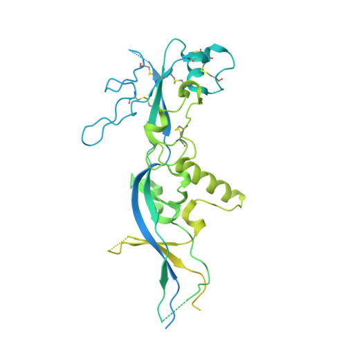
    5OW4

  • PubMed Abstract: 

    HAPLESS2 (HAP2) is a broadly conserved, gamete-expressed transmembrane protein that was shown recently to be structurally homologous to viral class II fusion proteins, which initiate fusion with host cells via insertion of fusion loops into the host membrane. However, the functional conformation of the HAP2 fusion loops has remained unknown, as the reported X-ray structure of Chlamydomonas reinhardtii HAP2 lacked this critical region. Here, we report a structure-guided alignment that reveals diversification of the proposed HAP2 fusion loops. Representative crystal structures show that in flowering plants, HAP2 has a single prominent fusion loop projecting an amphipathic helix at its apex, while in trypanosomes, three small nonpolar loops of HAP2 are poised to interact with the target membrane. A detailed structure-function analysis of the Arabidopsis HAP2 amphipathic fusion helix defines key residues that are essential for membrane insertion and for gamete fusion. Our study suggests that HAP2 may have evolved multiple modes of membrane insertion to accommodate the diversity of membrane environments it has encountered during eukaryotic evolution.


  • Organizational Affiliation

    Unité de Virologie Structurale, Institut Pasteur, Paris, France.


Macromolecules
Find similar proteins by:  (by identity cutoff)  |  3D Structure
Entity ID: 1
MoleculeChains Sequence LengthOrganismDetailsImage
Uncharacterized protein591Trypanosoma cruzi strain CL BrenerMutation(s): 0 
Gene Names: Tc00.1047053509105.4
UniProt
Find proteins for Q4DKJ2 (Trypanosoma cruzi (strain CL Brener))
Explore Q4DKJ2 
Go to UniProtKB:  Q4DKJ2
Entity Groups  
Sequence Clusters30% Identity50% Identity70% Identity90% Identity95% Identity100% Identity
UniProt GroupQ4DKJ2
Sequence Annotations
Expand
  • Reference Sequence
Experimental Data & Validation

Experimental Data

  • Method: X-RAY DIFFRACTION
  • Resolution: 3.10 Å
  • R-Value Free: 0.260 
  • R-Value Work: 0.233 
  • R-Value Observed: 0.234 
  • Space Group: P 61 2 2
Unit Cell:
Length ( Å )Angle ( ˚ )
a = 150.09α = 90
b = 150.09β = 90
c = 123.78γ = 120
Software Package:
Software NamePurpose
BUSTERrefinement
XDSdata reduction
XSCALEdata scaling
PHASERphasing

Structure Validation

View Full Validation Report


Entry History & Funding Information

Deposition Data


Funding OrganizationLocationGrant Number
European Research CouncilFrance340371 (CelCelFus)

Revision History  (Full details and data files)

  • Version 1.0: 2018-08-22
    Type: Initial release
  • Version 1.1: 2024-01-17
    Changes: Data collection, Database references, Refinement description