5OMK

R2-like ligand-binding oxidase with aerobically reconstituted metal cofactor before photoconversion


Experimental Data Snapshot

  • Method: X-RAY DIFFRACTION
  • Resolution: 1.70 Å
  • R-Value Free: 0.209 
  • R-Value Work: 0.178 
  • R-Value Observed: 0.180 

wwPDB Validation   3D Report Full Report


Ligand Structure Quality Assessment 


This is version 1.3 of the entry. See complete history


Literature

Driving Protein Conformational Changes with Light: Photoinduced Structural Rearrangement in a Heterobimetallic Oxidase.

Maugeri, P.T.Griese, J.J.Branca, R.M.Miller, E.K.Smith, Z.R.Eirich, J.Hogbom, M.Shafaat, H.S.

(2018) J Am Chem Soc 140: 1471-1480

  • DOI: https://doi.org/10.1021/jacs.7b11966
  • Primary Citation of Related Structures:  
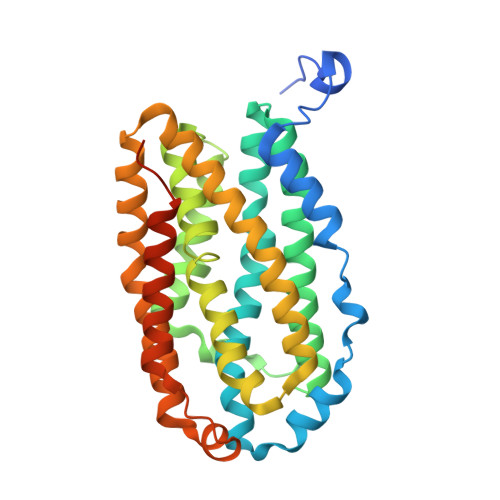
    5OMJ, 5OMK

  • PubMed Abstract: 

    The heterobimetallic R2lox protein binds both manganese and iron ions in a site-selective fashion and activates oxygen, ultimately performing C-H bond oxidation to generate a tyrosine-valine cross-link near the active site. In this work, we demonstrate that, following assembly, R2lox undergoes photoinduced changes to the active site geometry and metal coordination motif. Through spectroscopic, structural, and mass spectrometric characterization, the photoconverted species is found to consist of a tyrosinate-bound iron center following light-induced decarboxylation of a coordinating glutamate residue and cleavage of the tyrosine-valine cross-link. This process occurs with high quantum efficiencies (Φ = 3%) using violet and near-ultraviolet light, suggesting that the photodecarboxylation is initiated via ligand-to-metal charge transfer excitation. Site-directed mutagenesis and structural analysis suggest that the cross-linked tyrosine-162 is the coordinating residue. One primary product is observed following irradiation, indicating potential use of this class of proteins, which contains a putative substrate channel, for controlled photoinduced decarboxylation processes, with relevance for in vivo functionality of R2lox as well as application in environmental remediation.


  • Organizational Affiliation

    Biophysics Graduate Program, The Ohio State University , Columbus, Ohio 43210, United States.


Macromolecules
Find similar proteins by:  (by identity cutoff)  |  3D Structure
Entity ID: 1
MoleculeChains Sequence LengthOrganismDetailsImage
Ribonucleotide reductase small subunit316Geobacillus kaustophilus HTA426Mutation(s): 0 
Gene Names: GK2771
EC: 1.17.4.1
UniProt
Find proteins for Q5KW80 (Geobacillus kaustophilus (strain HTA426))
Explore Q5KW80 
Go to UniProtKB:  Q5KW80
Entity Groups  
Sequence Clusters30% Identity50% Identity70% Identity90% Identity95% Identity100% Identity
UniProt GroupQ5KW80
Sequence Annotations
Expand
  • Reference Sequence
Experimental Data & Validation

Experimental Data

  • Method: X-RAY DIFFRACTION
  • Resolution: 1.70 Å
  • R-Value Free: 0.209 
  • R-Value Work: 0.178 
  • R-Value Observed: 0.180 
  • Space Group: I 2 2 2
Unit Cell:
Length ( Å )Angle ( ˚ )
a = 55.666α = 90
b = 96.624β = 90
c = 128.399γ = 90
Software Package:
Software NamePurpose
PHENIXrefinement
XDSdata reduction
XDSdata scaling
PHENIXphasing

Structure Validation

View Full Validation Report


Ligand Structure Quality Assessment 


Entry History 

Deposition Data

Revision History  (Full details and data files)

  • Version 1.0: 2018-01-03
    Type: Initial release
  • Version 1.1: 2018-01-31
    Changes: Database references
  • Version 1.2: 2018-02-07
    Changes: Database references
  • Version 1.3: 2024-01-17
    Changes: Data collection, Database references, Refinement description