


Crystal structure of Omp36 from Enterobacter aerogenes
Ferrara, L., Naismith, J.To be published.
Experimental Data Snapshot
Entity ID: 1 | |||||
|---|---|---|---|---|---|
| Molecule | Chains | Sequence Length | Organism | Details | Image |
| Osmoporin Omp36 | A [auth C], B [auth A], C [auth D], D [auth B] | 354 | Klebsiella aerogenes | Mutation(s): 0 Gene Names: omp36 Membrane Entity: Yes | 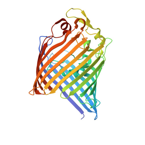 |
UniProt | |||||
Find proteins for Q9ALY0 (Klebsiella aerogenes) Explore Q9ALY0 Go to UniProtKB: Q9ALY0 | |||||
Entity Groups | |||||
| Sequence Clusters | 30% Identity50% Identity70% Identity90% Identity95% Identity100% Identity | ||||
| UniProt Group | Q9ALY0 | ||||
Sequence AnnotationsExpand | |||||
| |||||
| Ligands 1 Unique | |||||
|---|---|---|---|---|---|
| ID | Chains | Name / Formula / InChI Key | 2D Diagram | 3D Interactions | |
| C8E Query on C8E | E [auth A], F [auth D], G [auth B], H [auth B] | (HYDROXYETHYLOXY)TRI(ETHYLOXY)OCTANE C16 H34 O5 FEOZZFHAVXYAMB-UHFFFAOYSA-N |  | ||
| Length ( Å ) | Angle ( ˚ ) |
|---|---|
| a = 125.098 | α = 90 |
| b = 125.098 | β = 90 |
| c = 126.542 | γ = 120 |
| Software Name | Purpose |
|---|---|
| PHENIX | refinement |
| SCALA | data scaling |
| PHASER | phasing |
RCSB PDB (citation) is hosted by
RCSB PDB is a member of the

