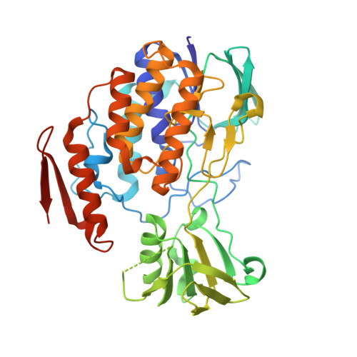
Crystal structure of pyrrolizidine alkaloid N-oxygenase from the grasshopper Zonocerus variegatus.
Kubitza, C., Faust, A., Gutt, M., Gath, L., Ober, D., Scheidig, A.J.(2018) Acta Crystallogr D Struct Biol 74: 422-432
- PubMed: 29717713
- DOI: https://doi.org/10.1107/S2059798318003510
- Primary Citation of Related Structures:
5NMW, 5NMX - PubMed Abstract:
The high-resolution crystal structure of the flavin-dependent monooxygenase (FMO) from the African locust Zonocerus variegatus is presented and the kinetics of structure-based protein variants are discussed. Z. variegatus expresses three flavin-dependent monooxygenase (ZvFMO) isoforms which contribute to a counterstrategy against pyrrolizidine alkaloids (PAs). PAs are protoxic compounds produced by some angiosperm lineages as a chemical defence against herbivores. N-Oxygenation of PAs and the accumulation of PA N-oxides within their haemolymph result in two evolutionary advantages for these insects: (i) they circumvent the defence mechanism of their food plants and (ii) they can use PA N-oxides to protect themselves against predators, which cannot cope with the toxic PAs. Despite a high degree of sequence identity and a similar substrate spectrum, the three ZvFMO isoforms differ greatly in enzyme activity. Here, the crystal structure of the Z. variegatus PA N-oxygenase (ZvPNO), the most active ZvFMO isoform, is reported at 1.6 Å resolution together with kinetic studies of a second isoform, ZvFMOa. This is the first available crystal structure of an FMO from class B (of six different FMO subclasses, A-F) within the family of flavin-dependent monooxygenases that originates from a more highly developed organism than yeast. Despite the differences in sequence between family members, their overall structure is very similar. This indicates the need for high conservation of the three-dimensional structure for this type of reaction throughout all kingdoms of life. Nevertheless, this structure provides the closest relative to the human enzyme that is currently available for modelling studies. Of note, the crystal structure of ZvPNO reveals a unique dimeric arrangement as well as small conformational changes within the active site that have not been observed before. A newly observed kink within helix α8 close to the substrate-binding path might indicate a potential mechanism for product release. The data show that even single amino-acid exchanges in the substrate-entry path, rather than the binding site, have a significant impact on the specific enzyme activity of the isoforms.
Organizational Affiliation:
Structural Biology, Zoological Institute, Kiel University, Am Botanischen Garten 1-9, 24118 Kiel, Germany.