


5NFF
Crystal structure of GP1 receptor binding domain from Morogoro virus
- PDB DOI: https://doi.org/10.2210/pdb5NFF/pdb
- Classification: VIRAL PROTEIN
- Organism(s): Morogoro mammarenavirus
- Expression System: Trichoplusia ni
- Mutation(s): No
- Deposited: 2017-03-14 Released: 2017-04-19
- Funding Organization(s): ISF - ICORE
Experimental Data Snapshot
- Method: X-RAY DIFFRACTION
- Resolution: 2.62 Å
- R-Value Free: 0.207
- R-Value Work: 0.174
- R-Value Observed: 0.176

This is version 2.1 of the entry. See complete history.

Macromolecules
Find similar proteins by:
(by identity cutoff) | 3D Structure
Entity ID: 1 | |||||
|---|---|---|---|---|---|
| Molecule | Chains | Sequence Length | Organism | Details | Image |
| Glycoprotein | 171 | Morogoro mammarenavirus | Mutation(s): 0 Gene Names: GPC | 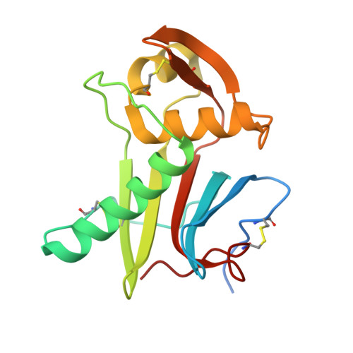 | |
UniProt | |||||
Find proteins for C6ZK00 (Morogoro mammarenavirus) Explore C6ZK00 Go to UniProtKB: C6ZK00 | |||||
Entity Groups | |||||
| Sequence Clusters | 30% Identity50% Identity70% Identity90% Identity95% Identity100% Identity | ||||
| UniProt Group | C6ZK00 | ||||
Sequence AnnotationsExpand | |||||
| |||||
Oligosaccharides
Entity ID: 2 | |||||
|---|---|---|---|---|---|
| Molecule | Chains | Length | 2D Diagram | Glycosylation | 3D Interactions |
| 2-acetamido-2-deoxy-beta-D-glucopyranose-(1-4)-2-acetamido-2-deoxy-beta-D-glucopyranose | Q, R | 2 |  | N-Glycosylation | |
Glycosylation Resources | |||||
GlyTouCan: G42666HT GlyCosmos: G42666HT GlyGen: G42666HT | |||||
Entity ID: 3 | |||||
|---|---|---|---|---|---|
| Molecule | Chains | Length | 2D Diagram | Glycosylation | 3D Interactions |
| alpha-D-mannopyranose-(1-6)-alpha-D-mannopyranose-(1-4)-2-acetamido-2-deoxy-beta-D-glucopyranose-(1-4)-2-acetamido-2-deoxy-beta-D-glucopyranose | S | 4 |  | N-Glycosylation | |
Glycosylation Resources | |||||
GlyTouCan: G04854NQ GlyCosmos: G04854NQ GlyGen: G04854NQ | |||||
Entity ID: 4 | |||||
|---|---|---|---|---|---|
| Molecule | Chains | Length | 2D Diagram | Glycosylation | 3D Interactions |
| alpha-D-mannopyranose-(1-2)-alpha-D-mannopyranose-(1-3)-alpha-D-mannopyranose-(1-4)-2-acetamido-2-deoxy-beta-D-glucopyranose-(1-4)-2-acetamido-2-deoxy-beta-D-glucopyranose | T | 5 |  | N-Glycosylation | |
Glycosylation Resources | |||||
GlyTouCan: G75436UI GlyCosmos: G75436UI GlyGen: G75436UI | |||||
Small Molecules
| Ligands 2 Unique | |||||
|---|---|---|---|---|---|
| ID | Chains | Name / Formula / InChI Key | 2D Diagram | 3D Interactions | |
| NAG Query on NAG | CA [auth D] DA [auth E] EA [auth E] HA [auth G] IA [auth G] | 2-acetamido-2-deoxy-beta-D-glucopyranose C8 H15 N O6 OVRNDRQMDRJTHS-FMDGEEDCSA-N |  | ||
| CIT Query on CIT | AA [auth C] BA [auth C] FA [auth E] GA [auth F] LA [auth I] | CITRIC ACID C6 H8 O7 KRKNYBCHXYNGOX-UHFFFAOYSA-N |  | ||
Experimental Data & Validation
Experimental Data
- Method: X-RAY DIFFRACTION
- Resolution: 2.62 Å
- R-Value Free: 0.207
- R-Value Work: 0.174
- R-Value Observed: 0.176
- Space Group: P 32
Unit Cell:
| Length ( Å ) | Angle ( ˚ ) |
|---|---|
| a = 127.769 | α = 90 |
| b = 127.769 | β = 90 |
| c = 251.704 | γ = 120 |
| Software Name | Purpose |
|---|---|
| PHENIX | refinement |
| PDB_EXTRACT | data extraction |
| XDS | data reduction |
| XSCALE | data scaling |
| PHASER | phasing |
Entry History & Funding Information
Deposition Data
- Released Date: 2017-04-19 Deposition Author(s): Israeli, H., Cohen-Dvashi, H., Shulman, A., Shimon, A., Diskin, R.
| Funding Organization | Location | Grant Number |
|---|---|---|
| ISF - ICORE | Israel | 1775/12 |
Revision History (Full details and data files)
- Version 1.0: 2017-04-19
Type: Initial release - Version 1.1: 2017-05-10
Changes: Database references - Version 1.2: 2019-08-14
Changes: Data collection - Version 2.0: 2020-07-29
Type: Remediation
Reason: Carbohydrate remediation
Changes: Advisory, Atomic model, Data collection, Derived calculations, Structure summary - Version 2.1: 2024-01-17
Changes: Data collection, Database references, Derived calculations, Refinement description, Structure summary
