5NF9

Structure of Galectin-3 CRD in complex with compound 2


Experimental Data Snapshot

  • Method: X-RAY DIFFRACTION
  • Resolution: 1.87 Å
  • R-Value Free: 0.214 
  • R-Value Work: 0.145 
  • R-Value Observed: 0.148 

wwPDB Validation   3D Report Full Report


This is version 1.1 of the entry. See complete history


Literature

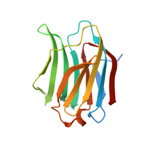
Biophysical and structural characterization of mono/di-arylated lactosamine derivatives interaction with human galectin-3.

Atmanene, C.Ronin, C.Teletchea, S.Gautier, F.M.Djedaini-Pilard, F.Ciesielski, F.Vivat, V.Grandjean, C.

(2017) Biochem Biophys Res Commun 489: 281-286

  • DOI: https://doi.org/10.1016/j.bbrc.2017.05.150
  • Primary Citation of Related Structures:  
    5NF7, 5NF9, 5NFA, 5NFB, 5NFC

  • PubMed Abstract: 

    Combination of biophysical and structural techniques allowed characterizing and uncovering the mechanisms underlying increased binding affinity of lactosamine derivatives for galectin 3. In particular, complementing information gathered from X-ray crystallography, native mass spectrometry and isothermal microcalorimetry showed favorable enthalpic contribution of cation-π interaction between lactosamine aryl substitutions and arginine residues from the carbohydrate recognition domain, which resulted in two log increase in compound binding affinity. This incrementing strategy allowed individual contribution of galectin inhibitor moieties to be dissected. Altogether, our results suggest that core and substituents of these saccharide-based inhibitors can be optimized separately, providing valuable tools to study the role of galectins in diseases.


  • Organizational Affiliation

    NovAliX Structural Biology Bioparc, Bd Sébastien Brant, BP30170, F-67405 Illkirch Cedex, France.


Macromolecules
Find similar proteins by:  (by identity cutoff)  |  3D Structure
Entity ID: 1
MoleculeChains Sequence LengthOrganismDetailsImage
Galectin-3147Homo sapiensMutation(s): 0 
Gene Names: LGALS3MAC2
UniProt & NIH Common Fund Data Resources
Find proteins for P17931 (Homo sapiens)
Explore P17931 
Go to UniProtKB:  P17931
PHAROS:  P17931
GTEx:  ENSG00000131981 
Entity Groups  
Sequence Clusters30% Identity50% Identity70% Identity90% Identity95% Identity100% Identity
UniProt GroupP17931
Sequence Annotations
Expand
  • Reference Sequence
Small Molecules
Ligands 1 Unique
IDChains Name / Formula / InChI Key2D Diagram3D Interactions
8VW
Query on 8VW

Download Ideal Coordinates CCD File 
B [auth A]~{N}-[(2~{R},3~{R},4~{R},5~{S},6~{R})-2-acetamido-6-(hydroxymethyl)-5-[(2~{S},3~{R},4~{S},5~{R},6~{R})-6-(hydroxymethyl)-3,4,5-tris(oxidanyl)oxan-2-yl]oxy-4-oxidanyl-oxan-3-yl]-3-methoxy-benzamide
C22 H32 N2 O12
SQNUIUJICOQQJY-NQSBSUMMSA-N
Binding Affinity Annotations 
IDSourceBinding Affinity
8VW Binding MOAD:  5NF9 Kd: 4700 (nM) from 1 assay(s)
Experimental Data & Validation

Experimental Data

  • Method: X-RAY DIFFRACTION
  • Resolution: 1.87 Å
  • R-Value Free: 0.214 
  • R-Value Work: 0.145 
  • R-Value Observed: 0.148 
  • Space Group: P 21 21 21
Unit Cell:
Length ( Å )Angle ( ˚ )
a = 36.87α = 90
b = 57.95β = 90
c = 62.98γ = 90
Software Package:
Software NamePurpose
REFMACrefinement
XDSdata reduction
XDSdata scaling
PHASERphasing

Structure Validation

View Full Validation Report


Entry History 

Deposition Data

Revision History  (Full details and data files)

  • Version 1.0: 2017-06-21
    Type: Initial release
  • Version 1.1: 2024-01-17
    Changes: Data collection, Database references, Refinement description