5NEV

CDK2/Cyclin A in complex with compound 73


Experimental Data Snapshot

  • Method: X-RAY DIFFRACTION
  • Resolution: 2.97 Å
  • R-Value Free: 0.261 
  • R-Value Work: 0.218 
  • R-Value Observed: 0.220 

wwPDB Validation   3D Report Full Report


Ligand Structure Quality Assessment 


This is version 1.1 of the entry. See complete history


Literature

Cyclin-Dependent Kinase (CDK) Inhibitors: Structure-Activity Relationships and Insights into the CDK-2 Selectivity of 6-Substituted 2-Arylaminopurines.

Coxon, C.R.Anscombe, E.Harnor, S.J.Martin, M.P.Carbain, B.Golding, B.T.Hardcastle, I.R.Harlow, L.K.Korolchuk, S.Matheson, C.J.Newell, D.R.Noble, M.E.Sivaprakasam, M.Tudhope, S.J.Turner, D.M.Wang, L.Z.Wedge, S.R.Wong, C.Griffin, R.J.Endicott, J.A.Cano, C.

(2017) J Med Chem 60: 1746-1767

  • DOI: https://doi.org/10.1021/acs.jmedchem.6b01254
  • Primary Citation of Related Structures:  
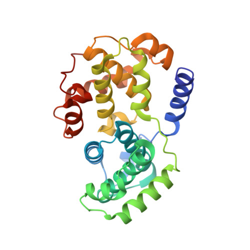
    5LQF, 5NEV

  • PubMed Abstract: 

    Purines and related heterocycles substituted at C-2 with 4'-sulfamoylanilino and at C-6 with a variety of groups have been synthesized with the aim of achieving selectivity of binding to CDK2 over CDK1. 6-Substituents that favor competitive inhibition at the ATP binding site of CDK2 were identified and typically exhibited 10-80-fold greater inhibition of CDK2 compared to CDK1. Most impressive was 4-((6-([1,1'-biphenyl]-3-yl)-9H-purin-2-yl)amino) benzenesulfonamide (73) that exhibited high potency toward CDK2 (IC 50 0.044 μM) but was ∼2000-fold less active toward CDK1 (IC 50 86 μM). This compound is therefore a useful tool for studies of cell cycle regulation. Crystal structures of inhibitor-kinase complexes showed that the inhibitor stabilizes a glycine-rich loop conformation that shapes the ATP ribose binding pocket and that is preferred in CDK2 but has not been observed in CDK1. This aspect of the active site may be exploited for the design of inhibitors that distinguish between CDK1 and CDK2.


  • Organizational Affiliation

    Newcastle Cancer Centre, Northern Institute for Cancer Research, School of Chemistry, Newcastle University , Bedson Building, Newcastle upon Tyne NE1 7RU, U.K.


Macromolecules
Find similar proteins by:  (by identity cutoff)  |  3D Structure
Entity ID: 1
MoleculeChains Sequence LengthOrganismDetailsImage
Cyclin-dependent kinase 2
A, C
303Homo sapiensMutation(s): 0 
Gene Names: CDK2CDKN2
EC: 2.7.11.22
UniProt & NIH Common Fund Data Resources
Find proteins for P24941 (Homo sapiens)
Explore P24941 
Go to UniProtKB:  P24941
PHAROS:  P24941
GTEx:  ENSG00000123374 
Entity Groups  
Sequence Clusters30% Identity50% Identity70% Identity90% Identity95% Identity100% Identity
UniProt GroupP24941
Sequence Annotations
Expand
  • Reference Sequence
Find similar proteins by:  (by identity cutoff)  |  3D Structure
Entity ID: 2
MoleculeChains Sequence LengthOrganismDetailsImage
Cyclin-A2
B, D
260Homo sapiensMutation(s): 0 
Gene Names: CCNA2CCN1CCNA
UniProt & NIH Common Fund Data Resources
Find proteins for P20248 (Homo sapiens)
Explore P20248 
Go to UniProtKB:  P20248
PHAROS:  P20248
GTEx:  ENSG00000145386 
Entity Groups  
Sequence Clusters30% Identity50% Identity70% Identity90% Identity95% Identity100% Identity
UniProt GroupP20248
Sequence Annotations
Expand
  • Reference Sequence
Small Molecules
Ligands 1 Unique
IDChains Name / Formula / InChI Key2D Diagram3D Interactions
72L
Query on 72L

Download Ideal Coordinates CCD File 
E [auth A],
F [auth C]
4-[[6-(3-phenylphenyl)-7~{H}-purin-2-yl]amino]benzenesulfonamide
C23 H18 N6 O2 S
FUGRWXRQJGJIER-UHFFFAOYSA-N
Modified Residues  1 Unique
IDChains TypeFormula2D DiagramParent
TPO
Query on TPO
A, C
L-PEPTIDE LINKINGC4 H10 N O6 PTHR
Experimental Data & Validation

Experimental Data

  • Method: X-RAY DIFFRACTION
  • Resolution: 2.97 Å
  • R-Value Free: 0.261 
  • R-Value Work: 0.218 
  • R-Value Observed: 0.220 
  • Space Group: P 21 21 21
Unit Cell:
Length ( Å )Angle ( ˚ )
a = 73.527α = 90
b = 132.053β = 90
c = 149.22γ = 90
Software Package:
Software NamePurpose
REFMACrefinement
XDSdata reduction
Aimlessdata scaling
PHASERphasing

Structure Validation

View Full Validation Report


Ligand Structure Quality Assessment 


Entry History 

Revision History  (Full details and data files)

  • Version 1.0: 2017-03-29
    Type: Initial release
  • Version 1.1: 2024-01-17
    Changes: Data collection, Database references, Refinement description