5MYC

Crystal structure of human 14-3-3 sigma in complex with LRRK2 peptide pS910


Experimental Data Snapshot

  • Method: X-RAY DIFFRACTION
  • Resolution: 1.46 Å
  • R-Value Free: 0.189 
  • R-Value Work: 0.166 
  • R-Value Observed: 0.167 

wwPDB Validation   3D Report Full Report


This is version 2.0 of the entry. See complete history


Literature

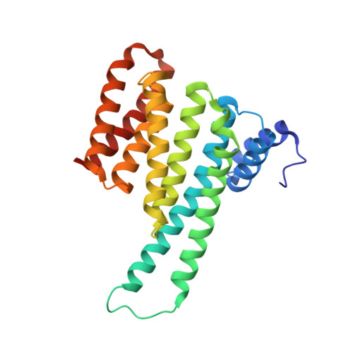
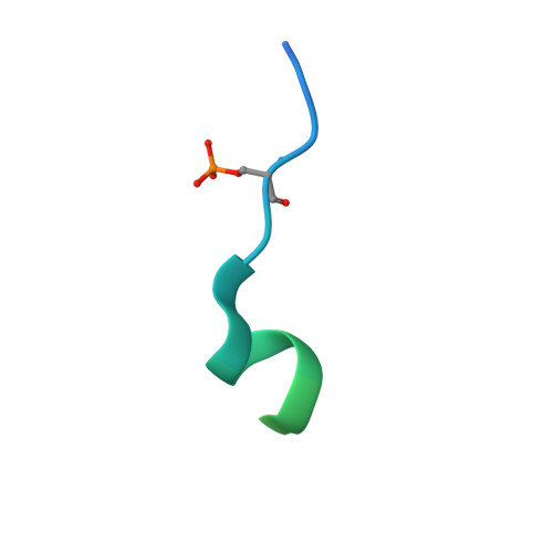
Structural interface between LRRK2 and 14-3-3 protein.

Stevers, L.M.de Vries, R.M.Doveston, R.G.Milroy, L.G.Brunsveld, L.Ottmann, C.

(2017) Biochem J 474: 1273-1287

  • DOI: https://doi.org/10.1042/BCJ20161078
  • Primary Citation of Related Structures:  
    5MY9, 5MYC

  • PubMed Abstract: 

    Binding of 14-3-3 proteins to leucine-rich repeat protein kinase 2 (LRRK2) is known to be impaired by many Parkinson's disease (PD)-relevant mutations. Abrogation of this interaction is connected to enhanced LRRK2 kinase activity, which in turn is implicated in increased ubiquitination of LRRK2, accumulation of LRRK2 into inclusion bodies and reduction in neurite length. Hence, the interaction between 14-3-3 and LRRK2 is of significant interest as a possible drug target for the treatment of PD. However, LRRK2 possesses multiple sites that, upon phosphorylation, can bind to 14-3-3, thus rendering the interaction relatively complex. Using biochemical assays and crystal structures, we characterize the multivalent interaction between these two proteins.


  • Organizational Affiliation

    Laboratory of Chemical Biology, Department of Biomedical Engineering and Institute for Complex Molecular Systems, Eindhoven University of Technology, PO Box 513, 5600 MB Eindhoven, The Netherlands.


Macromolecules
Find similar proteins by:  (by identity cutoff)  |  3D Structure
Entity ID: 1
MoleculeChains Sequence LengthOrganismDetailsImage
14-3-3 protein sigma236Homo sapiensMutation(s): 0 
Gene Names: SFNHME1
UniProt & NIH Common Fund Data Resources
Find proteins for P31947 (Homo sapiens)
Explore P31947 
Go to UniProtKB:  P31947
PHAROS:  P31947
GTEx:  ENSG00000175793 
Entity Groups  
Sequence Clusters30% Identity50% Identity70% Identity90% Identity95% Identity100% Identity
UniProt GroupP31947
Sequence Annotations
Expand
  • Reference Sequence
Find similar proteins by:  (by identity cutoff)  |  3D Structure
Entity ID: 2
MoleculeChains Sequence LengthOrganismDetailsImage
Leucine-rich repeat serine/threonine-protein kinase 2B [auth P]38Homo sapiensMutation(s): 0 
EC: 2.7.11.1
UniProt & NIH Common Fund Data Resources
Find proteins for Q5S007 (Homo sapiens)
Explore Q5S007 
Go to UniProtKB:  Q5S007
PHAROS:  Q5S007
GTEx:  ENSG00000188906 
Entity Groups  
Sequence Clusters30% Identity50% Identity70% Identity90% Identity95% Identity100% Identity
UniProt GroupQ5S007
Sequence Annotations
Expand
  • Reference Sequence
Experimental Data & Validation

Experimental Data

  • Method: X-RAY DIFFRACTION
  • Resolution: 1.46 Å
  • R-Value Free: 0.189 
  • R-Value Work: 0.166 
  • R-Value Observed: 0.167 
  • Space Group: C 2 2 21
Unit Cell:
Length ( Å )Angle ( ˚ )
a = 82.517α = 90
b = 112.411β = 90
c = 62.689γ = 90
Software Package:
Software NamePurpose
PHENIXrefinement
XDSdata reduction
XDSdata scaling

Structure Validation

View Full Validation Report


Entry History 

Deposition Data

Revision History  (Full details and data files)

  • Version 1.0: 2017-03-01
    Type: Initial release
  • Version 1.1: 2017-04-05
    Changes: Database references
  • Version 2.0: 2024-01-17
    Changes: Atomic model, Data collection, Database references, Derived calculations, Refinement description