5MWB

Human Notch-2 EGF11-13


Experimental Data Snapshot

  • Method: X-RAY DIFFRACTION
  • Resolution: 1.86 Å
  • R-Value Free: 0.264 
  • R-Value Work: 0.228 
  • R-Value Observed: 0.230 

wwPDB Validation   3D Report Full Report


Ligand Structure Quality Assessment 


This is version 2.1 of the entry. See complete history


Literature

Structural and functional dissection of the interplay between lipid and Notch binding by human Notch ligands.

Suckling, R.J.Korona, B.Whiteman, P.Chillakuri, C.Holt, L.Handford, P.A.Lea, S.M.

(2017) EMBO J 36: 2204-2215

  • DOI: https://doi.org/10.15252/embj.201796632
  • Primary Citation of Related Structures:  
    5MVX, 5MW5, 5MW7, 5MWB, 5MWF

  • PubMed Abstract: 

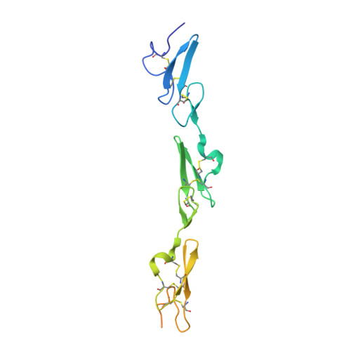
    Recent data have expanded our understanding of Notch signalling by identifying a C2 domain at the N-terminus of Notch ligands, which has both lipid- and receptor-binding properties. We present novel structures of human ligands Jagged2 and Delta-like4 and human Notch2, together with functional assays, which suggest that ligand-mediated coupling of membrane recognition and Notch binding is likely to be critical in establishing the optimal context for Notch signalling. Comparisons between the Jagged and Delta family show a huge diversity in the structures of the loops at the apex of the C2 domain implicated in membrane recognition and Jagged1 missense mutations, which affect these loops and are associated with extrahepatic biliary atresia, lead to a loss of membrane recognition, but do not alter Notch binding. Taken together, these data suggest that C2 domain binding to membranes is an important element in tuning ligand-dependent Notch signalling in different physiological contexts.


  • Organizational Affiliation

    Sir William Dunn School of Pathology, University of Oxford, Oxford, UK.


Macromolecules
Find similar proteins by:  (by identity cutoff)  |  3D Structure
Entity ID: 1
MoleculeChains Sequence LengthOrganismDetailsImage
Neurogenic locus notch homolog protein 2156Homo sapiensMutation(s): 0 
Gene Names: NOTCH2
UniProt & NIH Common Fund Data Resources
Find proteins for Q04721 (Homo sapiens)
Explore Q04721 
Go to UniProtKB:  Q04721
PHAROS:  Q04721
GTEx:  ENSG00000134250 
Entity Groups  
Sequence Clusters30% Identity50% Identity70% Identity90% Identity95% Identity100% Identity
UniProt GroupQ04721
Sequence Annotations
Expand
  • Reference Sequence
Oligosaccharides

Help

Entity ID: 2
MoleculeChains Length2D Diagram Glycosylation3D Interactions
beta-D-xylopyranose-(1-3)-beta-D-glucopyranose
B
2O-Glycosylation
Glycosylation Resources
GlyTouCan:  G61966PH
GlyCosmos:  G61966PH
GlyGen:  G61966PH
Experimental Data & Validation

Experimental Data

  • Method: X-RAY DIFFRACTION
  • Resolution: 1.86 Å
  • R-Value Free: 0.264 
  • R-Value Work: 0.228 
  • R-Value Observed: 0.230 
  • Space Group: P 21 21 21
Unit Cell:
Length ( Å )Angle ( ˚ )
a = 20.16α = 90
b = 49.79β = 90
c = 125.5γ = 90
Software Package:
Software NamePurpose
PHENIXrefinement
xia2data reduction
Aimlessdata scaling
PHASERphasing

Structure Validation

View Full Validation Report


Ligand Structure Quality Assessment 


Entry History & Funding Information

Deposition Data


Funding OrganizationLocationGrant Number
Medical Research Council (United Kingdom)United KingdomMR/L001187/1

Revision History  (Full details and data files)

  • Version 1.0: 2017-06-14
    Type: Initial release
  • Version 1.1: 2017-08-09
    Changes: Database references
  • Version 1.2: 2017-09-13
    Changes: Author supporting evidence
  • Version 2.0: 2020-07-29
    Type: Remediation
    Reason: Carbohydrate remediation
    Changes: Advisory, Atomic model, Data collection, Derived calculations, Structure summary
  • Version 2.1: 2024-01-17
    Changes: Data collection, Database references, Derived calculations, Refinement description, Structure summary