5MVT

Crystal structure of an A-DNA dodecamer featuring an alternating pyrimidine-purine sequence


Experimental Data Snapshot

  • Method: X-RAY DIFFRACTION
  • Resolution: 1.90 Å
  • R-Value Free: 0.225 
  • R-Value Work: 0.202 
  • R-Value Observed: 0.203 

wwPDB Validation   3D Report Full Report


This is version 1.6 of the entry. See complete history


Literature

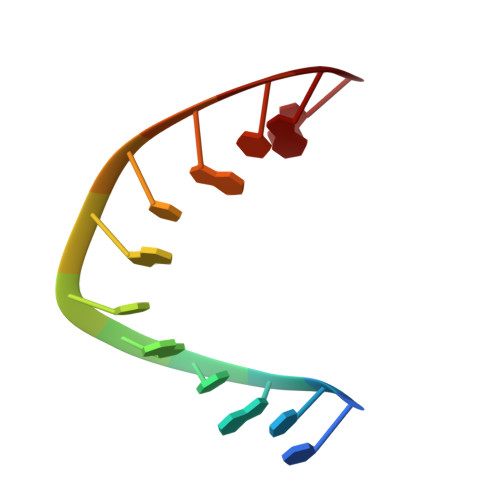
5-Formylcytosine does not change the global structure of DNA.

Hardwick, J.S.Ptchelkine, D.El-Sagheer, A.H.Tear, I.Singleton, D.Phillips, S.E.V.Lane, A.N.Brown, T.

(2017) Nat Struct Mol Biol 24: 544-552

  • DOI: https://doi.org/10.1038/nsmb.3411
  • Primary Citation of Related Structures:  
    5MVK, 5MVL, 5MVP, 5MVQ, 5MVT, 5MVU

  • PubMed Abstract: 

    The mechanism by which the recently identified DNA modification 5-formylcytosine ( f C) is recognized by epigenetic writer and reader proteins is not known. Recently, an unusual DNA structure, F-DNA, has been proposed as the basis for enzyme recognition of clusters of f C. We used NMR and X-ray crystallography to compare several modified DNA duplexes with unmodified analogs and found that in the crystal state the duplexes all belong to the A family, whereas in solution they are all members of the B family. We found that, contrary to previous findings, f C does not significantly affect the structure of DNA, although there are modest local differences at the modification sites. Hence, global conformation changes are unlikely to account for the recognition of this modified base, and our structural data favor a mechanism that operates at base-pair resolution for the recognition of f C by epigenome-modifying enzymes.


  • Organizational Affiliation

    Department of Chemistry, University of Oxford, Chemistry Research Laboratory, Oxford, UK.


Macromolecules

Find similar nucleic acids by:  Sequence   |   3D Structure  

Entity ID: 1
MoleculeChains LengthOrganismImage
DNA
A, B
12synthetic construct
Sequence Annotations
Expand
  • Reference Sequence
Small Molecules
Ligands 1 Unique
IDChains Name / Formula / InChI Key2D Diagram3D Interactions
3CO
Query on 3CO

Download Ideal Coordinates CCD File 
C [auth B]COBALT (III) ION
Co
JAWGVVJVYSANRY-UHFFFAOYSA-N
Experimental Data & Validation

Experimental Data

  • Method: X-RAY DIFFRACTION
  • Resolution: 1.90 Å
  • R-Value Free: 0.225 
  • R-Value Work: 0.202 
  • R-Value Observed: 0.203 
  • Space Group: P 32 2 1
Unit Cell:
Length ( Å )Angle ( ˚ )
a = 44.288α = 90
b = 44.288β = 90
c = 57.841γ = 120
Software Package:
Software NamePurpose
PHENIXrefinement
XDSdata reduction
Aimlessdata scaling
PHASERphasing

Structure Validation

View Full Validation Report


Entry History & Funding Information

Deposition Data


Funding OrganizationLocationGrant Number
Biotechnology and Biological Sciences Research CouncilBB/J001694/2

Revision History  (Full details and data files)

  • Version 1.0: 2017-05-10
    Type: Initial release
  • Version 1.1: 2017-05-17
    Changes: Database references
  • Version 1.2: 2017-05-24
    Changes: Database references
  • Version 1.3: 2017-06-14
    Changes: Database references
  • Version 1.4: 2017-08-30
    Changes: Author supporting evidence
  • Version 1.5: 2019-10-16
    Changes: Data collection
  • Version 1.6: 2024-01-17
    Changes: Data collection, Database references, Refinement description