5MVN

X-ray structure of the M205W mutant of GLIC in complex with propofol


Experimental Data Snapshot

  • Method: X-RAY DIFFRACTION
  • Resolution: 3.49 Å
  • R-Value Free: 0.232 
  • R-Value Work: 0.218 
  • R-Value Observed: 0.218 

wwPDB Validation   3D Report Full Report


Ligand Structure Quality Assessment 


This is version 1.3 of the entry. See complete history


Literature

Structural Basis for a Bimodal Allosteric Mechanism of General Anesthetic Modulation in Pentameric Ligand-Gated Ion Channels.

Fourati, Z.Howard, R.J.Heusser, S.A.Hu, H.Ruza, R.R.Sauguet, L.Lindahl, E.Delarue, M.

(2018) Cell Rep 23: 993-1004

  • DOI: https://doi.org/10.1016/j.celrep.2018.03.108
  • Primary Citation of Related Structures:  
    5MUO, 5MUR, 5MVN, 5MZQ, 5NKJ, 6EMX

  • PubMed Abstract: 

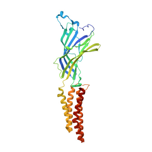
    Ion channel modulation by general anesthetics is a vital pharmacological process with implications for receptor biophysics and drug development. Functional studies have implicated conserved sites of both potentiation and inhibition in pentameric ligand-gated ion channels, but a detailed structural mechanism for these bimodal effects is lacking. The prokaryotic model protein GLIC recapitulates anesthetic modulation of human ion channels, and it is accessible to structure determination in both apparent open and closed states. Here, we report ten X-ray structures and electrophysiological characterization of GLIC variants in the presence and absence of general anesthetics, including the surgical agent propofol. We show that general anesthetics can allosterically favor closed channels by binding in the pore or favor open channels via various subsites in the transmembrane domain. Our results support an integrated, multi-site mechanism for allosteric modulation, and they provide atomic details of both potentiation and inhibition by one of the most common general anesthetics.


  • Organizational Affiliation

    Unit of Structural Dynamics of Macromolecules, Institut Pasteur and UMR 3528 du CNRS, 75015 Paris, France.


Macromolecules
Find similar proteins by:  (by identity cutoff)  |  3D Structure
Entity ID: 1
MoleculeChains Sequence LengthOrganismDetailsImage
Proton-gated ion channel
A, B, C, D, E
317Gloeobacter violaceus PCC 7421Mutation(s): 1 
Gene Names: glvIglr4197
Membrane Entity: Yes 
UniProt
Find proteins for Q7NDN8 (Gloeobacter violaceus (strain ATCC 29082 / PCC 7421))
Explore Q7NDN8 
Go to UniProtKB:  Q7NDN8
Entity Groups  
Sequence Clusters30% Identity50% Identity70% Identity90% Identity95% Identity100% Identity
UniProt GroupQ7NDN8
Sequence Annotations
Expand
  • Reference Sequence
Small Molecules
Ligands 6 Unique
IDChains Name / Formula / InChI Key2D Diagram3D Interactions
PLC
Query on PLC

Download Ideal Coordinates CCD File 
CA [auth C]
DA [auth C]
G [auth A]
H [auth A]
I [auth A]
CA [auth C],
DA [auth C],
G [auth A],
H [auth A],
I [auth A],
JA [auth D],
LA [auth D],
MA [auth D],
NA [auth D],
R [auth A],
T [auth B],
U [auth B],
UA [auth E],
V [auth B],
VA [auth E]
DIUNDECYL PHOSPHATIDYL CHOLINE
C32 H65 N O8 P
IJFVSSZAOYLHEE-SSEXGKCCSA-O
LMT
Query on LMT

Download Ideal Coordinates CCD File 
HA [auth C]
P [auth A]
Q [auth A]
RA [auth D]
Z [auth B]
HA [auth C],
P [auth A],
Q [auth A],
RA [auth D],
Z [auth B],
ZA [auth E]
DODECYL-BETA-D-MALTOSIDE
C24 H46 O11
NLEBIOOXCVAHBD-QKMCSOCLSA-N
PFL
Query on PFL

Download Ideal Coordinates CCD File 
AB [auth E],
BA [auth B],
IA [auth C],
SA [auth D]
2,6-BIS(1-METHYLETHYL)PHENOL
C12 H18 O
OLBCVFGFOZPWHH-UHFFFAOYSA-N
ACT
Query on ACT

Download Ideal Coordinates CCD File 
AA [auth B]
F [auth A]
GA [auth C]
KA [auth D]
O [auth A]
AA [auth B],
F [auth A],
GA [auth C],
KA [auth D],
O [auth A],
QA [auth D],
S [auth B],
TA [auth E],
Y [auth B],
YA [auth E]
ACETATE ION
C2 H3 O2
QTBSBXVTEAMEQO-UHFFFAOYSA-M
CL
Query on CL

Download Ideal Coordinates CCD File 
EA [auth C]
J [auth A]
K [auth A]
L [auth A]
OA [auth D]
EA [auth C],
J [auth A],
K [auth A],
L [auth A],
OA [auth D],
W [auth B],
WA [auth E]
CHLORIDE ION
Cl
VEXZGXHMUGYJMC-UHFFFAOYSA-M
NA
Query on NA

Download Ideal Coordinates CCD File 
FA [auth C]
M [auth A]
N [auth A]
PA [auth D]
X [auth B]
FA [auth C],
M [auth A],
N [auth A],
PA [auth D],
X [auth B],
XA [auth E]
SODIUM ION
Na
FKNQFGJONOIPTF-UHFFFAOYSA-N
Experimental Data & Validation

Experimental Data

  • Method: X-RAY DIFFRACTION
  • Resolution: 3.49 Å
  • R-Value Free: 0.232 
  • R-Value Work: 0.218 
  • R-Value Observed: 0.218 
  • Space Group: C 1 2 1
Unit Cell:
Length ( Å )Angle ( ˚ )
a = 181.229α = 90
b = 132.474β = 102.5
c = 159.759γ = 90
Software Package:
Software NamePurpose
BUSTERrefinement
XDSdata reduction
Aimlessdata scaling
REFMACphasing

Structure Validation

View Full Validation Report


Ligand Structure Quality Assessment 


Entry History 

Deposition Data

Revision History  (Full details and data files)

  • Version 1.0: 2018-02-14
    Type: Initial release
  • Version 1.1: 2018-06-20
    Changes: Data collection, Database references
  • Version 1.2: 2019-10-16
    Changes: Data collection
  • Version 1.3: 2024-01-17
    Changes: Data collection, Database references, Refinement description