5MV9

Structure of human Myosin 7a C-terminal MyTH4-FERM domain in complex with harmonin-a PDZ3 domain


Experimental Data Snapshot

  • Method: X-RAY DIFFRACTION
  • Resolution: 2.60 Å
  • R-Value Free: 0.237 
  • R-Value Work: 0.180 
  • R-Value Observed: 0.182 

wwPDB Validation   3D Report Full Report


This is version 2.0 of the entry. See complete history


Literature

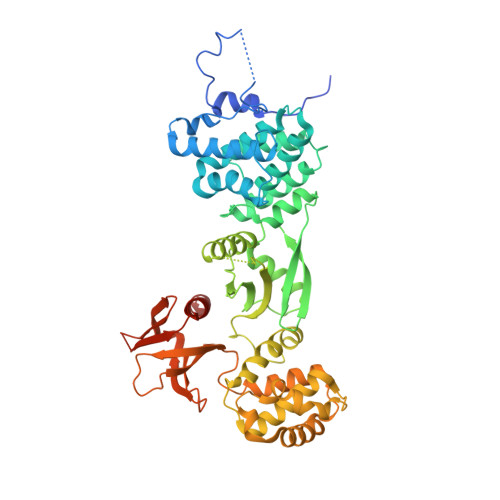
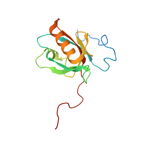
Myosin 7 and its adaptors link cadherins to actin.

Yu, I.M.Planelles-Herrero, V.J.Sourigues, Y.Moussaoui, D.Sirkia, H.Kikuti, C.Stroebel, D.Titus, M.A.Houdusse, A.

(2017) Nat Commun 8: 15864-15864

  • DOI: https://doi.org/10.1038/ncomms15864
  • Primary Citation of Related Structures:  
    5MV7, 5MV8, 5MV9

  • PubMed Abstract: 

    Cadherin linkages between adjacent stereocilia and microvilli are essential for mechanotransduction and maintaining their organization. They are anchored to actin through interaction of their cytoplasmic domains with related tripartite complexes consisting of a class VII myosin and adaptor proteins: Myo7a/SANS/Harmonin in stereocilia and Myo7b/ANKS4B/Harmonin in microvilli. Here, we determine high-resolution structures of Myo7a and Myo7b C-terminal MyTH4-FERM domain (MF2) and unveil how they recognize harmonin using a novel binding mode. Systematic definition of interactions between domains of the tripartite complex elucidates how the complex assembles and prevents possible self-association of harmonin-a. Several Myo7a deafness mutants that map to the surface of MF2 disrupt harmonin binding, revealing the molecular basis for how they impact the formation of the tripartite complex and disrupt mechanotransduction. Our results also suggest how switching between different harmonin isoforms can regulate the formation of networks with Myo7a motors and coordinate force sensing in stereocilia.


  • Organizational Affiliation

    Structural Motility, Institut Curie, PSL Research University, CNRS, UMR 144, F-75005 Paris, France.


Macromolecules
Find similar proteins by:  (by identity cutoff)  |  3D Structure
Entity ID: 1
MoleculeChains Sequence LengthOrganismDetailsImage
Unconventional myosin-VIIa527Homo sapiensMutation(s): 0 
Gene Names: MYO7AUSH1B
UniProt & NIH Common Fund Data Resources
Find proteins for Q13402 (Homo sapiens)
Explore Q13402 
Go to UniProtKB:  Q13402
PHAROS:  Q13402
GTEx:  ENSG00000137474 
Entity Groups  
Sequence Clusters30% Identity50% Identity70% Identity90% Identity95% Identity100% Identity
UniProt GroupQ13402
Sequence Annotations
Expand
  • Reference Sequence
Find similar proteins by:  (by identity cutoff)  |  3D Structure
Entity ID: 2
MoleculeChains Sequence LengthOrganismDetailsImage
cDNA FLJ51329, highly similar to Harmonin130Homo sapiensMutation(s): 0 
UniProt & NIH Common Fund Data Resources
Find proteins for Q9Y6N9 (Homo sapiens)
Explore Q9Y6N9 
Go to UniProtKB:  Q9Y6N9
PHAROS:  Q9Y6N9
GTEx:  ENSG00000006611 
Entity Groups  
Sequence Clusters30% Identity50% Identity70% Identity90% Identity95% Identity100% Identity
UniProt GroupQ9Y6N9
Sequence Annotations
Expand
  • Reference Sequence
Experimental Data & Validation

Experimental Data

  • Method: X-RAY DIFFRACTION
  • Resolution: 2.60 Å
  • R-Value Free: 0.237 
  • R-Value Work: 0.180 
  • R-Value Observed: 0.182 
  • Space Group: C 2 2 21
Unit Cell:
Length ( Å )Angle ( ˚ )
a = 121.52α = 90
b = 151.71β = 90
c = 100.8γ = 90
Software Package:
Software NamePurpose
BUSTERrefinement
XDSdata reduction
XDSdata scaling
PHASERphasing

Structure Validation

View Full Validation Report


Entry History 

Deposition Data

Revision History  (Full details and data files)

  • Version 1.0: 2017-07-05
    Type: Initial release
  • Version 1.1: 2017-07-12
    Changes: Database references
  • Version 2.0: 2024-01-17
    Changes: Atomic model, Data collection, Database references, Refinement description