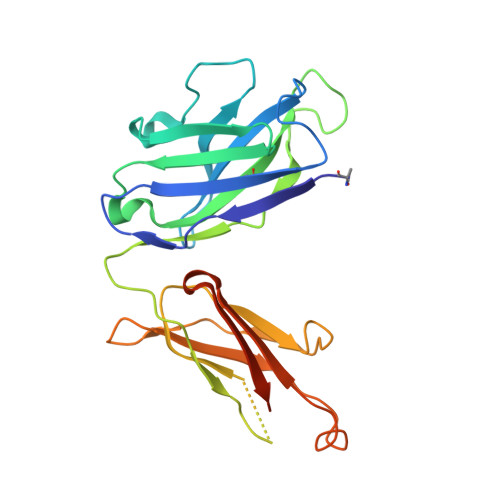
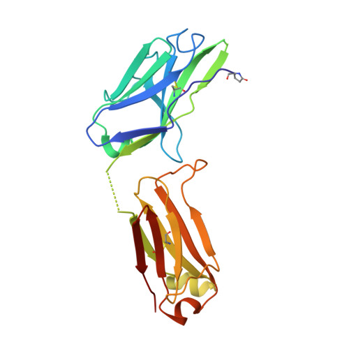
Structure and Recognition of a Novel HIV-1 gp120-gp41 Interface Antibody that Caused MPER Exposure through Viral Escape.
Wibmer, C.K., Gorman, J., Ozorowski, G., Bhiman, J.N., Sheward, D.J., Elliott, D.H., Rouelle, J., Smira, A., Joyce, M.G., Ndabambi, N., Druz, A., Asokan, M., Burton, D.R., Connors, M., Abdool Karim, S.S., Mascola, J.R., Robinson, J.E., Ward, A.B., Williamson, C., Kwong, P.D., Morris, L., Moore, P.L.(2017) PLoS Pathog 13: e1006074-e1006074
- PubMed: 28076415
- DOI: https://doi.org/10.1371/journal.ppat.1006074
- Primary Citation of Related Structures:
5F89, 5MP6 - PubMed Abstract:
A comprehensive understanding of the regions on HIV-1 envelope trimers targeted by broadly neutralizing antibodies may contribute to rational design of an HIV-1 vaccine. We previously identified a participant in the CAPRISA cohort, CAP248, who developed trimer-specific antibodies capable of neutralizing 60% of heterologous viruses at three years post-infection. Here, we report the isolation by B cell culture of monoclonal antibody CAP248-2B, which targets a novel membrane proximal epitope including elements of gp120 and gp41. Despite low maximum inhibition plateaus, often below 50% inhibitory concentrations, the breadth of CAP248-2B significantly correlated with donor plasma. Site-directed mutagenesis, X-ray crystallography, and negative-stain electron microscopy 3D reconstructions revealed how CAP248-2B recognizes a cleavage-dependent epitope that includes the gp120 C terminus. While this epitope is distinct, it overlapped in parts of gp41 with the epitopes of broadly neutralizing antibodies PGT151, VRC34, 35O22, 3BC315, and 10E8. CAP248-2B has a conformationally variable paratope with an unusually long 19 amino acid light chain third complementarity determining region. Two phenylalanines at the loop apex were predicted by docking and mutagenesis data to interact with the viral membrane. Neutralization by CAP248-2B is not dependent on any single glycan proximal to its epitope, and low neutralization plateaus could not be completely explained by N- or O-linked glycosylation pathway inhibitors, furin co-transfection, or pre-incubation with soluble CD4. Viral escape from CAP248-2B involved a cluster of rare mutations in the gp120-gp41 cleavage sites. Simultaneous introduction of these mutations into heterologous viruses abrogated neutralization by CAP248-2B, but enhanced neutralization sensitivity to 35O22, 4E10, and 10E8 by 10-100-fold. Altogether, this study expands the region of the HIV-1 gp120-gp41 quaternary interface that is a target for broadly neutralizing antibodies and identifies a set of mutations in the gp120 C terminus that exposes the membrane-proximal external region of gp41, with potential utility in HIV vaccine design.
Organizational Affiliation:
Centre for HIV and STIs, National Institute for Communicable Diseases (NICD), of the National Health Laboratory Service (NHLS), Johannesburg, South Africa.