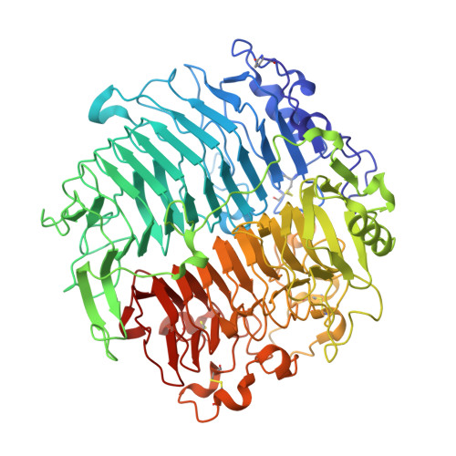
5M5Z

Chaetomium thermophilum beta-1-3-glucanase


Experimental Data Snapshot

  • Method: X-RAY DIFFRACTION
  • Resolution: 1.25 Å
  • R-Value Free: 0.141 
  • R-Value Work: 0.123 
  • R-Value Observed: 0.123 

wwPDB Validation   3D Report Full Report


Ligand Structure Quality Assessment 


This is version 3.1 of the entry. See complete history


Literature

Crystal structure and biological implications of a glycoside hydrolase family 55 beta-1,3-glucanase from Chaetomium thermophilum.

Papageorgiou, A.C.Chen, J.Li, D.

(2017) Biochim Biophys Acta 1865: 1030-1038

  • DOI: https://doi.org/10.1016/j.bbapap.2017.05.002
  • Primary Citation of Related Structures:  
    5M5Z, 5M60

  • PubMed Abstract: 

    Crystal structures of a β-1,3-glucanase from the thermophilic fungus Chaetomium thermophilum were determined at 1.20 and 1.42Å resolution in the free and glucose-bound form, respectively. This is the third structure of a family 55 glycoside hydrolase (GH55) member and the second from a fungus. Based on comparative structural studies and site-directed mutagenesis, Glu654 is proposed as the catalytic acid residue. The substrate binding cleft exhibits restricted access on one side, rendering the enzyme as an exo-β-1,3-glucanase as confirmed also by thin layer chromatography experiments. A lack of stacking interactions was found at the substrate binding cleft, suggesting that interactions at positions -1, +1 and +2 are sufficient to orientate the substrate. A binding pocket was identified that could explain binding of branched laminarin and accumulation of laminaritriose.


  • Organizational Affiliation

    Turku Centre for Biotechnology, University of Turku and Åbo Akademi University, 20521 Turku, Finland. Electronic address: tassos.papageorgiou@btk.fi.


Macromolecules
Find similar proteins by:  (by identity cutoff)  |  3D Structure
Entity ID: 1
MoleculeChains Sequence LengthOrganismDetailsImage
Beta-1,3-glucanase757Thermochaetoides thermophilaMutation(s): 0 
Gene Names: gluN
UniProt
Find proteins for G0S3N2 (Chaetomium thermophilum (strain DSM 1495 / CBS 144.50 / IMI 039719))
Explore G0S3N2 
Go to UniProtKB:  G0S3N2
Entity Groups  
Sequence Clusters30% Identity50% Identity70% Identity90% Identity95% Identity100% Identity
UniProt GroupG0S3N2
Sequence Annotations
Expand
  • Reference Sequence
Oligosaccharides

Help

Entity ID: 2
MoleculeChains Length2D Diagram Glycosylation3D Interactions
2-acetamido-2-deoxy-beta-D-glucopyranose-(1-4)-2-acetamido-2-deoxy-beta-D-glucopyranose
B, C
2N-Glycosylation
Glycosylation Resources
GlyTouCan:  G42666HT
GlyCosmos:  G42666HT
GlyGen:  G42666HT
Experimental Data & Validation

Experimental Data

  • Method: X-RAY DIFFRACTION
  • Resolution: 1.25 Å
  • R-Value Free: 0.141 
  • R-Value Work: 0.123 
  • R-Value Observed: 0.123 
  • Space Group: P 1 21 1
Unit Cell:
Length ( Å )Angle ( ˚ )
a = 63.774α = 90
b = 85.277β = 92.83
c = 66.765γ = 90
Software Package:
Software NamePurpose
PHENIXrefinement
XDSdata reduction
Aimlessdata scaling
PHASERphasing

Structure Validation

View Full Validation Report


Ligand Structure Quality Assessment 


Entry History 

Deposition Data

Revision History  (Full details and data files)

  • Version 1.0: 2017-05-17
    Type: Initial release
  • Version 1.1: 2017-06-14
    Changes: Database references
  • Version 2.0: 2020-03-11
    Changes: Atomic model, Data collection, Derived calculations, Polymer sequence
  • Version 3.0: 2020-07-29
    Type: Remediation
    Reason: Carbohydrate remediation
    Changes: Advisory, Atomic model, Data collection, Derived calculations, Structure summary
  • Version 3.1: 2024-01-17
    Changes: Data collection, Database references, Refinement description, Structure summary