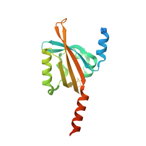
5LUV

Short LOV protein W619_1 in apo-state


Experimental Data Snapshot

  • Method: X-RAY DIFFRACTION
  • Resolution: 2.50 Å
  • R-Value Free: 0.253 
  • R-Value Work: 0.211 
  • R-Value Observed: 0.213 

wwPDB Validation   3D Report Full Report


This is version 1.2 of the entry. See complete history


Literature

Structure of a LOV protein in apo-state and implications for construction of LOV-based optical tools.

Arinkin, V.Granzin, J.Rollen, K.Krauss, U.Jaeger, K.E.Willbold, D.Batra-Safferling, R.

(2017) Sci Rep 7: 42971-42971

  • DOI: https://doi.org/10.1038/srep42971
  • Primary Citation of Related Structures:  
    5LUV

  • PubMed Abstract: 

    Unique features of Light-Oxygen-Voltage (LOV) proteins like relatively small size (~12-19 kDa), inherent modularity, highly-tunable photocycle and oxygen-independent fluorescence have lately been exploited for the generation of optical tools. Structures of LOV domains reported so far contain a flavin chromophore per protein molecule. Here we report two new findings on the short LOV protein W619_1-LOV from Pseudomonas putida. First, the apo-state crystal structure of W619_1-LOV at 2.5 Å resolution reveals conformational rearrangements in the secondary structure elements lining the chromophore pocket including elongation of the Fα helix, shortening of the Eα-Fα loop and partial unfolding of the Eα helix. Second, the apo W619_1-LOV protein binds both natural and structurally modified flavin chromophores. Remarkably different photophysical and photochemical properties of W619_1-LOV bound to 7-methyl-8-chloro-riboflavin (8-Cl-RF) and lumichrome imply application of these variants as novel optical tools as they offer advantages such as no adduct state formation, and a broader choice of wavelengths for in vitro studies.


  • Organizational Affiliation

    Institute of Complex Systems, ICS-6: Structural Biochemistry, Forschungszentrum Jülich, 52425 Jülich, Germany.


Macromolecules
Find similar proteins by:  (by identity cutoff)  |  3D Structure
Entity ID: 1
MoleculeChains Sequence LengthOrganismDetailsImage
Putative PAS/PAC sensor protein
A, B
166Pseudomonas putida W619Mutation(s): 0 
Gene Names: PputW619_0812
UniProt
Find proteins for B1J385 (Pseudomonas putida (strain W619))
Explore B1J385 
Go to UniProtKB:  B1J385
Entity Groups  
Sequence Clusters30% Identity50% Identity70% Identity90% Identity95% Identity100% Identity
UniProt GroupB1J385
Sequence Annotations
Expand
  • Reference Sequence
Experimental Data & Validation

Experimental Data

  • Method: X-RAY DIFFRACTION
  • Resolution: 2.50 Å
  • R-Value Free: 0.253 
  • R-Value Work: 0.211 
  • R-Value Observed: 0.213 
  • Space Group: I 41
Unit Cell:
Length ( Å )Angle ( ˚ )
a = 109.538α = 90
b = 109.538β = 90
c = 95.209γ = 90
Software Package:
Software NamePurpose
PHENIXrefinement
XDSdata reduction
Aimlessdata scaling
MOLREPphasing

Structure Validation

View Full Validation Report


Entry History & Funding Information

Deposition Data


Funding OrganizationLocationGrant Number
Bundesministerium fuer Bildung und ForschungGermany031A167B

Revision History  (Full details and data files)

  • Version 1.0: 2017-02-22
    Type: Initial release
  • Version 1.1: 2017-03-01
    Changes: Database references
  • Version 1.2: 2024-01-17
    Changes: Data collection, Database references, Refinement description