5LS7

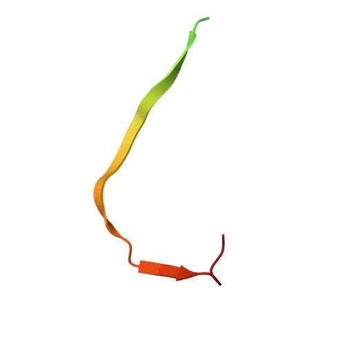
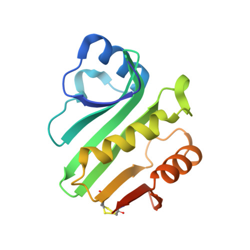
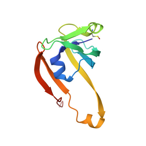
Complex of wild type E. coli alpha aspartate decarboxylase with its processing factor PanZ


Experimental Data Snapshot

  • Method: X-RAY DIFFRACTION
  • Resolution: 1.16 Å
  • R-Value Free: 0.137 
  • R-Value Work: 0.113 
  • R-Value Observed: 0.115 

wwPDB Validation   3D Report Full Report


Ligand Structure Quality Assessment 


This is version 2.1 of the entry. See complete history


Literature

The Mechanism of Regulation of Pantothenate Biosynthesis by the PanD-PanZAcCoA Complex Reveals an Additional Mode of Action for the Antimetabolite N-Pentyl Pantothenamide (N5-Pan).

Arnott, Z.L.P.Nozaki, S.Monteiro, D.C.F.Morgan, H.E.Pearson, A.R.Niki, H.Webb, M.E.

(2017) Biochemistry 56: 4931-4939

  • DOI: https://doi.org/10.1021/acs.biochem.7b00509
  • Primary Citation of Related Structures:  
    5LS7

  • PubMed Abstract: 

    The antimetabolite pentyl pantothenamide has broad spectrum antibiotic activity but exhibits enhanced activity against Escherichia coli. The PanDZ complex has been proposed to regulate the pantothenate biosynthetic pathway in E. coli by limiting the supply of β-alanine in response to coenzyme A concentration. We show that formation of such a complex between activated aspartate decarboxylase (PanD) and PanZ leads to sequestration of the pyruvoyl cofactor as a ketone hydrate and demonstrate that both PanZ overexpression-linked β-alanine auxotrophy and pentyl pantothenamide toxicity are due to formation of this complex. This both demonstrates that the PanDZ complex regulates pantothenate biosynthesis in a cellular context and validates the complex as a target for antibiotic development.


  • Organizational Affiliation

    Astbury Centre for Structural Molecular Biology and School of Chemistry, University of Leeds , Leeds LS2 9JT, U.K.


Macromolecules
Find similar proteins by:  (by identity cutoff)  |  3D Structure
Entity ID: 1
MoleculeChains Sequence LengthOrganismDetailsImage
Aspartate 1-decarboxylase41Escherichia coli K-12Mutation(s): 0 
Gene Names: panDb0131JW0127
EC: 4.1.1.11
UniProt
Find proteins for P0A790 (Escherichia coli (strain K12))
Explore P0A790 
Go to UniProtKB:  P0A790
Entity Groups  
Sequence Clusters30% Identity50% Identity70% Identity90% Identity95% Identity100% Identity
UniProt GroupP0A790
Sequence Annotations
Expand
  • Reference Sequence
Find similar proteins by:  (by identity cutoff)  |  3D Structure
Entity ID: 2
MoleculeChains Sequence LengthOrganismDetailsImage
PanD maturation factor137Escherichia coli K-12Mutation(s): 0 
Gene Names: panMpanZyhhKb3459JW3424
UniProt
Find proteins for P37613 (Escherichia coli (strain K12))
Explore P37613 
Go to UniProtKB:  P37613
Entity Groups  
Sequence Clusters30% Identity50% Identity70% Identity90% Identity95% Identity100% Identity
UniProt GroupP37613
Sequence Annotations
Expand
  • Reference Sequence
Find similar proteins by:  (by identity cutoff)  |  3D Structure
Entity ID: 3
MoleculeChains Sequence LengthOrganismDetailsImage
Aspartate 1-decarboxylaseC [auth D]102Escherichia coli K-12Mutation(s): 0 
Gene Names: panDb0131JW0127
EC: 4.1.1.11
UniProt
Find proteins for P0A790 (Escherichia coli (strain K12))
Explore P0A790 
Go to UniProtKB:  P0A790
Entity Groups  
Sequence Clusters30% Identity50% Identity70% Identity90% Identity95% Identity100% Identity
UniProt GroupP0A790
Sequence Annotations
Expand
  • Reference Sequence
Small Molecules
Ligands 7 Unique
IDChains Name / Formula / InChI Key2D Diagram3D Interactions
ACO
Query on ACO

Download Ideal Coordinates CCD File 
F [auth B]ACETYL COENZYME *A
C23 H38 N7 O17 P3 S
ZSLZBFCDCINBPY-ZSJPKINUSA-N
PEG
Query on PEG

Download Ideal Coordinates CCD File 
E [auth B],
M [auth D]
DI(HYDROXYETHYL)ETHER
C4 H10 O3
MTHSVFCYNBDYFN-UHFFFAOYSA-N
GOL
Query on GOL

Download Ideal Coordinates CCD File 
D [auth B],
J [auth D]
GLYCEROL
C3 H8 O3
PEDCQBHIVMGVHV-UHFFFAOYSA-N
SCN
Query on SCN

Download Ideal Coordinates CCD File 
K [auth D],
L [auth D]
THIOCYANATE ION
C N S
ZMZDMBWJUHKJPS-UHFFFAOYSA-M
CO2
Query on CO2

Download Ideal Coordinates CCD File 
H [auth B],
I [auth B],
O [auth D]
CARBON DIOXIDE
C O2
CURLTUGMZLYLDI-UHFFFAOYSA-N
MG
Query on MG

Download Ideal Coordinates CCD File 
G [auth B]MAGNESIUM ION
Mg
JLVVSXFLKOJNIY-UHFFFAOYSA-N
74C
Query on 74C

Download Ideal Coordinates CCD File 
N [auth D]methyl radical
C H3
VNWKTOKETHGBQD-UHFFFAOYSA-N
Modified Residues  1 Unique
IDChains TypeFormula2D DiagramParent
CSO
Query on CSO
C [auth D]L-PEPTIDE LINKINGC3 H7 N O3 SCYS
Binding Affinity Annotations 
IDSourceBinding Affinity
ACO Binding MOAD:  5LS7 Kd: 524 (nM) from 1 assay(s)
Experimental Data & Validation

Experimental Data

  • Method: X-RAY DIFFRACTION
  • Resolution: 1.16 Å
  • R-Value Free: 0.137 
  • R-Value Work: 0.113 
  • R-Value Observed: 0.115 
  • Space Group: I 4
Unit Cell:
Length ( Å )Angle ( ˚ )
a = 85.863α = 90
b = 85.863β = 90
c = 80.055γ = 90
Software Package:
Software NamePurpose
REFMACrefinement
XDSdata reduction
Aimlessdata scaling
MOLREPphasing

Structure Validation

View Full Validation Report


Ligand Structure Quality Assessment 


Entry History & Funding Information

Deposition Data


Funding OrganizationLocationGrant Number
Wellcome TrustUnited Kingdom096684/Z/11/Z
Wellcome TrustUnited Kingdom105615/Z/14/Z
German Research FoundationGermanyExcellence Cluster: Hamburg Centre for Ultrafast Imaging
Wellcome TrustUnited Kingdom094232/Z/10/Z

Revision History  (Full details and data files)

  • Version 1.0: 2017-09-13
    Type: Initial release
  • Version 1.1: 2017-09-20
    Changes: Database references
  • Version 1.2: 2017-09-27
    Changes: Database references
  • Version 2.0: 2023-11-15
    Changes: Atomic model, Data collection, Database references, Derived calculations
  • Version 2.1: 2024-01-17
    Changes: Refinement description