5LRD

Crystal structure of Glycogen Phosphorylase b in complex with KS242


Experimental Data Snapshot

  • Method: X-RAY DIFFRACTION
  • Resolution: 1.80 Å
  • R-Value Free: 0.187 
  • R-Value Work: 0.160 
  • R-Value Observed: 0.162 

wwPDB Validation   3D Report Full Report


Ligand Structure Quality Assessment 


This is version 1.1 of the entry. See complete history


Literature

van der Waals interactions govern C-beta-d-glucopyranosyl triazoles' nM inhibitory potency in human liver glycogen phosphorylase.

Kantsadi, A.L.Stravodimos, G.A.Kyriakis, E.Chatzileontiadou, D.S.M.Solovou, T.G.A.Kun, S.Bokor, E.Somsak, L.Leonidas, D.D.

(2017) J Struct Biol 199: 57-67

  • DOI: https://doi.org/10.1016/j.jsb.2017.05.001
  • Primary Citation of Related Structures:  
    5LRC, 5LRD, 5LRF

  • PubMed Abstract: 

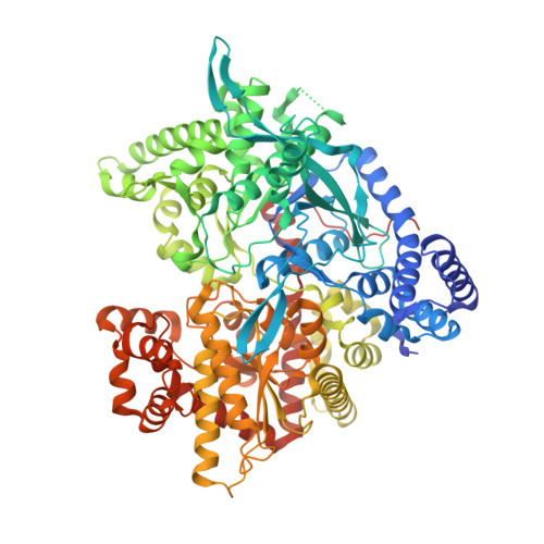
    3-(C-Glucopyranosyl)-5aryl-1,2,4-triazoles with an aryl moiety larger than phenyl have been shown to have strong inhibitory potency (K i values in the range of upper nM) for human liver glycogen phosphorylase (hlGP), a pharmacologically relevant target for diabetes type 2. In this study we investigate in a comparative manner the inhibitory effect of the above triazoles and their respective imidazoles on hlGPa. Kinetic studies show that the imidazole derivatives are 6-8 times more potent than their corresponding triazoles. We also seek to answer how the type of the aryl moiety affects the potency in hlGPa, and by determination of the crystal structure of rmGPb in complex with the triazole derivatives the structural basis of their inhibitory efficacy is also elucidated. Our studies revealed that the van der Waals interactions between the aryl moiety and residues in a hydrophobic pocket within the active site are mainly responsible for the variations in the potency of these inhibitors.


  • Organizational Affiliation

    Department of Biochemistry and Biotechnology, University of Thessaly, Biopolis, 41500 Larissa, Greece.


Macromolecules
Find similar proteins by:  (by identity cutoff)  |  3D Structure
Entity ID: 1
MoleculeChains Sequence LengthOrganismDetailsImage
Glycogen phosphorylase, muscle form843Oryctolagus cuniculusMutation(s): 0 
EC: 2.4.1.1
UniProt
Find proteins for P00489 (Oryctolagus cuniculus)
Explore P00489 
Go to UniProtKB:  P00489
Entity Groups  
Sequence Clusters30% Identity50% Identity70% Identity90% Identity95% Identity100% Identity
UniProt GroupP00489
Sequence Annotations
Expand
  • Reference Sequence
Small Molecules
Ligands 3 Unique
IDChains Name / Formula / InChI Key2D Diagram3D Interactions
KS2
Query on KS2

Download Ideal Coordinates CCD File 
B [auth A](2~{R},3~{S},4~{R},5~{R},6~{S})-2-(hydroxymethyl)-6-[5-(4-methylphenyl)-4~{H}-1,2,4-triazol-3-yl]oxane-3,4,5-triol
C15 H19 N3 O5
YAGKTBWCGRXONX-UJPOAAIJSA-N
PLP
Query on PLP

Download Ideal Coordinates CCD File 
C [auth A]PYRIDOXAL-5'-PHOSPHATE
C8 H10 N O6 P
NGVDGCNFYWLIFO-UHFFFAOYSA-N
DMS
Query on DMS

Download Ideal Coordinates CCD File 
D [auth A],
E [auth A]
DIMETHYL SULFOXIDE
C2 H6 O S
IAZDPXIOMUYVGZ-UHFFFAOYSA-N
Binding Affinity Annotations 
IDSourceBinding Affinity
KS2 Binding MOAD:  5LRD Ki: 1700 (nM) from 1 assay(s)
Experimental Data & Validation

Experimental Data

  • Method: X-RAY DIFFRACTION
  • Resolution: 1.80 Å
  • R-Value Free: 0.187 
  • R-Value Work: 0.160 
  • R-Value Observed: 0.162 
  • Space Group: P 43 21 2
Unit Cell:
Length ( Å )Angle ( ˚ )
a = 128.54α = 90
b = 128.54β = 90
c = 116.3γ = 90
Software Package:
Software NamePurpose
REFMACrefinement
MOSFLMdata reduction
SCALAdata scaling
MOSFLMdata reduction

Structure Validation

View Full Validation Report


Ligand Structure Quality Assessment 


Entry History 

Deposition Data

Revision History  (Full details and data files)

  • Version 1.0: 2017-06-14
    Type: Initial release
  • Version 1.1: 2017-06-28
    Changes: Database references