


X-ray structure of uridine phosphorylase from Vibrio cholerae in complex with cytidine and cytosine at 1.11 A resolution
Prokofev, I.I., Gabdoulkhakov, A.G., Lashkov, A.A., Balaev, V.V., Betzel, C., Mikhailov, A.M.To be published.
Experimental Data Snapshot
Entity ID: 1 | |||||
|---|---|---|---|---|---|
| Molecule | Chains | Sequence Length | Organism | Details | Image |
| Uridine phosphorylase | 253 | Vibrio cholerae | Mutation(s): 0 Gene Names: udp, udp_1, DN30_1909 EC: 2.4.2.3 | 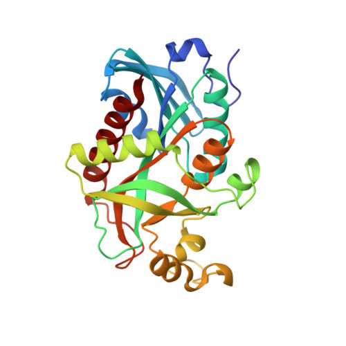 | |
UniProt | |||||
Find proteins for Q9K4U1 (Vibrio cholerae) Explore Q9K4U1 Go to UniProtKB: Q9K4U1 | |||||
Entity Groups | |||||
| Sequence Clusters | 30% Identity50% Identity70% Identity90% Identity95% Identity100% Identity | ||||
| UniProt Group | Q9K4U1 | ||||
Sequence AnnotationsExpand | |||||
| |||||
| Ligands 7 Unique | |||||
|---|---|---|---|---|---|
| ID | Chains | Name / Formula / InChI Key | 2D Diagram | 3D Interactions | |
| CTN Query on CTN | FA [auth E] I [auth A] JA [auth F] P [auth B] U [auth C] | 4-AMINO-1-BETA-D-RIBOFURANOSYL-2(1H)-PYRIMIDINONE C9 H13 N3 O5 UHDGCWIWMRVCDJ-XVFCMESISA-N |  | ||
| CYT Query on CYT | GA [auth E] J [auth A] KA [auth F] Q [auth B] V [auth C] | 6-AMINOPYRIMIDIN-2(1H)-ONE C4 H5 N3 O OPTASPLRGRRNAP-UHFFFAOYSA-N |  | ||
| SO4 Query on SO4 | L [auth A] | SULFATE ION O4 S QAOWNCQODCNURD-UHFFFAOYSA-L |  | ||
| GOL Query on GOL | AA [auth D] HA [auth E] K [auth A] LA [auth F] R [auth B] | GLYCEROL C3 H8 O3 PEDCQBHIVMGVHV-UHFFFAOYSA-N |  | ||
| EDO Query on EDO | DA [auth E], G [auth A], O [auth B] | 1,2-ETHANEDIOL C2 H6 O2 LYCAIKOWRPUZTN-UHFFFAOYSA-N |  | ||
| K Query on K | EA [auth E], H [auth A], T [auth C] | POTASSIUM ION K NPYPAHLBTDXSSS-UHFFFAOYSA-N |  | ||
| CL Query on CL | BA [auth E] CA [auth E] IA [auth F] M [auth B] N [auth B] | CHLORIDE ION Cl VEXZGXHMUGYJMC-UHFFFAOYSA-M |  | ||
| Length ( Å ) | Angle ( ˚ ) |
|---|---|
| a = 64.258 | α = 69.35 |
| b = 71.772 | β = 72.17 |
| c = 89.228 | γ = 85.78 |
| Software Name | Purpose |
|---|---|
| PHENIX | refinement |
| XSCALE | data scaling |
| MOLREP | phasing |
| PDB_EXTRACT | data extraction |
| XDS | data reduction |
| Funding Organization | Location | Grant Number |
|---|---|---|
| RFBR | Russian Federation | 14-04-00952a |
| Grant of President of Russian Federation | Russian Federation | MK-9246.2016.3 |
RCSB PDB (citation) is hosted by
RCSB PDB is a member of the

