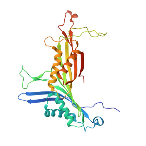
Catalysis and Structure of Zebrafish Urate Oxidase Provide Insights into the Origin of Hyperuricemia in Hominoids.
Marchetti, M., Liuzzi, A., Fermi, B., Corsini, R., Folli, C., Speranzini, V., Gandolfi, F., Bettati, S., Ronda, L., Cendron, L., Berni, R., Zanotti, G., Percudani, R.(2016) Sci Rep 6: 38302-38302
- PubMed: 27922051
- DOI: https://doi.org/10.1038/srep38302
- Primary Citation of Related Structures:
5LL1, 5M98 - PubMed Abstract:
Urate oxidase (Uox) catalyses the first reaction of oxidative uricolysis, a three-step enzymatic pathway that allows some animals to eliminate purine nitrogen through a water-soluble compound. Inactivation of the pathway in hominoids leads to elevated levels of sparingly soluble urate and puts humans at risk of hyperuricemia and gout. The uricolytic activities lost during evolution can be replaced by enzyme therapy. Here we report on the functional and structural characterization of Uox from zebrafish and the effects on the enzyme of the missense mutation (F216S) that preceded Uox pseudogenization in hominoids. Using a kinetic assay based on the enzymatic suppression of the spectroscopic interference of the Uox reaction product, we found that the F216S mutant has the same turnover number of the wild-type enzyme but a much-reduced affinity for the urate substrate and xanthine inhibitor. Our results indicate that the last functioning Uox in hominoid evolution had an increased Michaelis constant, possibly near to upper end of the normal range of urate in the human serum (~300 μM). Changes in the renal handling of urate during primate evolution can explain the genetic modification of uricolytic activities in the hominoid lineage without the need of assuming fixation of deleterious mutations.
Organizational Affiliation:
Department of Life Sciences, University of Parma, 43124, Parma, Italy.