5L35

Cryo-EM structure of bacteriophage Sf6 at 2.9 Angstrom resolution

  • Classification: VIRUS
  • Organism(s): Lederbergvirus Sf6
  • Mutation(s): No 

  • Deposited: 2016-08-03 Released: 2017-03-22 
  • Deposition Author(s): Zhao, H., Tang, L.
  • Funding Organization(s): National Institutes of Health/National Institute of General Medical Sciences (NIH/NIGMS)

Experimental Data Snapshot

  • Method: ELECTRON MICROSCOPY
  • Resolution: 2.89 Å
  • Aggregation State: PARTICLE 
  • Reconstruction Method: SINGLE PARTICLE 

wwPDB Validation   3D Report Full Report


This is version 1.5 of the entry. See complete history


Literature

Structure of a headful DNA-packaging bacterial virus at 2.9 angstrom resolution by electron cryo-microscopy.

Zhao, H.Li, K.Lynn, A.Y.Aron, K.E.Yu, G.Jiang, W.Tang, L.

(2017) Proc Natl Acad Sci U S A 114: 3601-3606

  • DOI: https://doi.org/10.1073/pnas.1615025114
  • Primary Citation of Related Structures:  
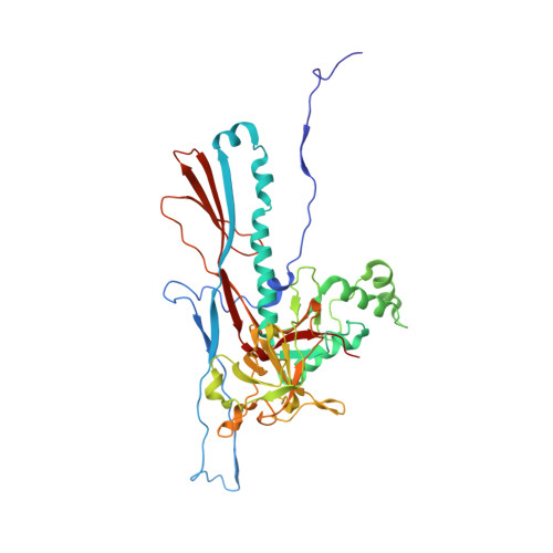
    5L35

  • PubMed Abstract: 

    The enormous prevalence of tailed DNA bacteriophages on this planet is enabled by highly efficient self-assembly of hundreds of protein subunits into highly stable capsids. These capsids can stand with an internal pressure as high as ∼50 atmospheres as a result of the phage DNA-packaging process. Here we report the complete atomic model of the headful DNA-packaging bacteriophage Sf6 at 2.9 Å resolution determined by electron cryo-microscopy. The structure reveals the DNA-inflated, tensed state of a robust protein shell assembled via noncovalent interactions. Remarkable global conformational polymorphism of capsid proteins, a network formed by extended N arms, mortise-and-tenon-like intercapsomer joints, and abundant β-sheet-like mainchain:mainchain intermolecular interactions, confers significant strength yet also flexibility required for capsid assembly and DNA packaging. Differential formations of the hexon and penton are mediated by a drastic α-helix-to-β-strand structural transition. The assembly scheme revealed here may be common among tailed DNA phages and herpesviruses.


  • Organizational Affiliation

    Department of Molecular Biosciences, University of Kansas, Lawrence, KS 66045.


Macromolecules
Find similar proteins by:  (by identity cutoff)  |  3D Structure
Entity ID: 1
MoleculeChains Sequence LengthOrganismDetailsImage
Gene 5 protein
A, B, C, D, E
A, B, C, D, E, F, G
422Lederbergvirus Sf6Mutation(s): 0 
UniProt
Find proteins for Q716H0 (Shigella phage Sf6)
Explore Q716H0 
Go to UniProtKB:  Q716H0
Entity Groups  
Sequence Clusters30% Identity50% Identity70% Identity90% Identity95% Identity100% Identity
UniProt GroupQ716H0
Sequence Annotations
Expand
  • Reference Sequence
Small Molecules
Ligands 1 Unique
IDChains Name / Formula / InChI Key2D Diagram3D Interactions
CL
Query on CL

Download Ideal Coordinates CCD File 
H [auth E]CHLORIDE ION
Cl
VEXZGXHMUGYJMC-UHFFFAOYSA-M
Experimental Data & Validation

Experimental Data

  • Method: ELECTRON MICROSCOPY
  • Resolution: 2.89 Å
  • Aggregation State: PARTICLE 
  • Reconstruction Method: SINGLE PARTICLE 
EM Software:
TaskSoftware PackageVersion
RECONSTRUCTIONjspr1
MODEL REFINEMENTPHENIX1.10

Structure Validation

View Full Validation Report


Entry History & Funding Information

Deposition Data


Funding OrganizationLocationGrant Number
National Institutes of Health/National Institute of General Medical Sciences (NIH/NIGMS)United StatesGM090010

Revision History  (Full details and data files)

  • Version 1.0: 2017-03-22
    Type: Initial release
  • Version 1.1: 2017-04-05
    Changes: Database references
  • Version 1.2: 2017-04-19
    Changes: Database references
  • Version 1.3: 2018-07-18
    Changes: Data collection
  • Version 1.4: 2019-11-27
    Changes: Author supporting evidence
  • Version 1.5: 2024-03-06
    Changes: Data collection, Database references, Derived calculations