5L0P

Symmetry-based assembly of a two-dimensional protein lattice


Experimental Data Snapshot

  • Method: X-RAY DIFFRACTION
  • Resolution: 2.30 Å
  • R-Value Free: 0.236 
  • R-Value Work: 0.198 

wwPDB Validation   3D Report Full Report


This is version 1.3 of the entry. See complete history


Literature

Symmetry based assembly of a 2 dimensional protein lattice.

Poulos, S.Agah, S.Jallah, N.Faham, S.

(2017) PLoS One 12: e0174485-e0174485

  • DOI: https://doi.org/10.1371/journal.pone.0174485
  • Primary Citation of Related Structures:  
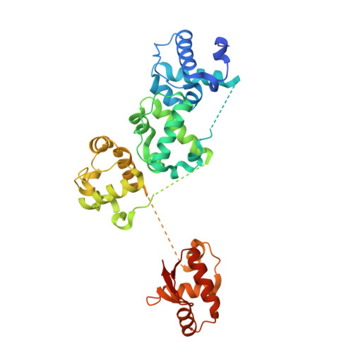
    5L0P

  • PubMed Abstract: 

    The design of proteins that self-assemble into higher order architectures is of great interest due to their potential application in nanotechnology. Specifically, the self-assembly of proteins into ordered lattices is of special interest to the field of structural biology. Here we designed a 2 dimensional (2D) protein lattice using a fusion of a tandem repeat of three TelSAM domains (TTT) to the Ferric uptake regulator (FUR) domain. We determined the structure of the designed (TTT-FUR) fusion protein to 2.3 Å by X-ray crystallographic methods. In agreement with the design, a 2D lattice composed of TelSAM fibers interdigitated by the FUR domain was observed. As expected, the fusion of a tandem repeat of three TelSAM domains formed 21 screw axis, and the self-assembly of the ordered oligomer was under pH control. We demonstrated that the fusion of TTT to a domain having a 2-fold symmetry, such as the FUR domain, can produce an ordered 2D lattice. The TTT-FUR system combines features from the rotational symmetry matching approach with the oligomer driven crystallization method. This TTT-FUR fusion was amenable to X-ray crystallographic methods, and is a promising crystallization chaperone.


  • Organizational Affiliation

    Department of Molecular Physiology and Biological Physics, University of Virginia School of Medicine, Charlottesville, Virginia, United States.


Macromolecules
Find similar proteins by:  (by identity cutoff)  |  3D Structure
Entity ID: 1
MoleculeChains Sequence LengthOrganismDetailsImage
Transcription factor ETV6, Transcription factor ETV6, Transcription factor ETV6, Ferric uptake regulation protein chimera353Homo sapiensKlebsiella pneumoniaeMutation(s): 0 
Gene Names: ETV6TELTEL1fur
UniProt & NIH Common Fund Data Resources
Find proteins for P41212 (Homo sapiens)
Explore P41212 
Go to UniProtKB:  P41212
PHAROS:  P41212
GTEx:  ENSG00000139083 
Find proteins for P45599 (Klebsiella pneumoniae)
Explore P45599 
Go to UniProtKB:  P45599
Entity Groups  
Sequence Clusters30% Identity50% Identity70% Identity90% Identity95% Identity100% Identity
UniProt GroupsP41212P45599
Sequence Annotations
Expand
  • Reference Sequence
Experimental Data & Validation

Experimental Data

  • Method: X-RAY DIFFRACTION
  • Resolution: 2.30 Å
  • R-Value Free: 0.236 
  • R-Value Work: 0.198 
  • Space Group: C 1 2 1
Unit Cell:
Length ( Å )Angle ( ˚ )
a = 131.313α = 90
b = 54.833β = 128.47
c = 88.48γ = 90
Software Package:
Software NamePurpose
REFMACrefinement
HKL-2000data reduction
HKL-2000data scaling
PHASERphasing

Structure Validation

View Full Validation Report


Entry History 

Deposition Data

  • Released Date: 2017-03-29 
  • Deposition Author(s): Faham, S.

Revision History  (Full details and data files)

  • Version 1.0: 2017-03-29
    Type: Initial release
  • Version 1.1: 2017-05-03
    Changes: Database references
  • Version 1.2: 2017-08-16
    Changes: Refinement description
  • Version 1.3: 2023-10-04
    Changes: Data collection, Database references, Refinement description